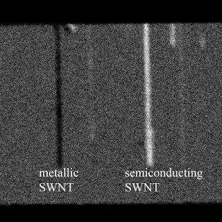
WEST LAFAYETTE, Ind. - Researchers have demonstrated a new imaging tool for rapidly screening structures called single-wall carbon nanotubes, possibly hastening their use in creating a new class of computers and electronics that are faster and consume less power than today's.
The semiconducting nanostructures might be used to revolutionize electronics by replacing conventional silicon components and circuits. However, one obstacle in their application is that metallic versions form unavoidably during the manufacturing process, contaminating the semiconducting nanotubes.
Now researchers have discovered that an advanced imaging technology could solve this problem, said Ji-Xin Cheng, an associate professor of biomedical engineering and chemistry at Purdue University.
"The imaging system uses a pulsing laser to deposit energy into the nanotubes, pumping the nanotubes from a ground state to an excited state," he said. "Then, another laser called a probe senses the excited nanotubes and reveals the contrast between metallic and semiconductor tubes."
"When you make nanocircuits, you only want the semiconducting ones, so it's very important to have a method to identify the metallic nanotubes," Yang said.
The paper was written by Purdue physics doctoral student Yookyung Jung; biomedical engineering research scientist Mikhail N. Slipchenko; Chang-Hua Liu, an electrical engineering graduate student at the University of Michigan; Alexander E. Ribbe, manager of the Nanotechnology Group in Purdue's Department of Chemistry; Zhaohui Zhong, an assistant professor of electrical engineering and computer science at Michigan; and Yang and Cheng. The Michigan researchers produced the nanotubes.
Semiconductors such as silicon conduct electricity under some conditions but not others, making them ideal for controlling electrical current in devices such as transistors and diodes.
The nanotubes have a diameter of about 1 nanometer, or roughly the length of 10 hydrogen atoms strung together, making them far too small to be seen with a conventional light microscope.
"They can be seen with an atomic force microscope, but this only tells you the morphology and surface features, not the metallic state of the nanotube," Cheng said.
The transient absorption imaging technique represents the only rapid method for telling the difference between the two types of nanotubes. The technique is "label free," meaning it does not require that the nanotubes be marked with dyes, making it potentially practical for manufacturing, he said.
The researchers performed the technique with nanotubes placed on a glass surface. Future work will focus on performing the imaging when nanotubes are on a silicon surface to determine how well it would work in industrial applications.
"We have begun this work on a silicon substrate, and preliminary results are very good," Cheng said.
Future research also may study how electrons travel inside individual nanotubes. ###
The research is funded by the National Science Foundation.
Related websites:
Ji-Xin Cheng: engineering.purdue.edu/BME/Research/Labs/Cheng Weldon School of Biomedical Engineering: www.purdue.edu/bme Department of Chemistry: www.chem.purdue.edu/ Chen Yang: www.chem.purdue.edu/people/faculty/faculty
Contact: Emil Venere venere@purdue.edu 765-494-4709 Purdue University


No comments:
Post a Comment