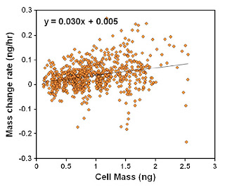
CHAMPAIGN, Ill. — University of Illinois researchers are using a new kind of microsensor to answer one of the weightiest questions in biology – the relationship between cell mass and growth rate.
The team, led by electrical and computer engineering and bioengineering professor Rashid Bashir, published its results in the online early edition of the Proceedings of the National Academy of Sciences.
"It's merging micro-scale engineering and cell biology," said Bashir, who also directs the Micro and Nanotechnology Engineering Laboratory at Illinois. "We can help advance biology by fabricating new tools that can be used to address important questions in cell biology, cancer research and tissue engineering."
The mechanics of cellular growth and division are important not only for basic biology, but also for diagnostics, drug development, tissue engineering and understanding cancer. For example, documenting these processes could help identify specific drug targets to slow or stop the uncontrolled growth of cancer cells.
"As you make the structure smaller and smaller, it becomes more sensitive to the mass that's placed on it," Bashir said. "A cell is a few nanograms in mass or smaller. If we can make our sensor small enough, then it becomes sensitive to cell mass."
The researchers developed an array of hundreds of sensors on a chip. They can culture cells on the chip similar to the way scientists grow cells in a dish. Thus, they can collect data from many cells at once, while still recording individual cellular measurements.
Another advantage of these microsensors is the ability to image cells with microscopes while cells grow on the sensors. Researchers can track the cells visually, opening the possibilities of tracking various cellular processes in conjunction with changes in mass.
"Imaging acts as a control. You can actually watch the cell divide and grow and correlate that to your measurements. It really validates what you have," Bashir said. "There are lots of optical measurements that now you can integrate with mass sensing."
Through measurements of live and fixed cells, the researchers were also able to extract physical properties such as stiffness through mathematical modeling. Some cell types are stiffer than others; for example, bone cells are more firm, while neurons are more gelatinous. Mechanical science and engineering professors Narayana Aluru and K. Jimmy Hsia, co-authors of the paper, performed extensive analytical and numerical simulation to reveal how cell stiffness and contact area affect mass measurement.
Next, the researchers plan to extend the study to other cell lines, and explore more optical measurements and fluorescent markers.
"These technologies can also be used for diagnostic purposes, or for screening. For example, we could study cell growth and mass and changes in the cell structure based on drugs or chemicals," Bashir said. ###
The National Science Foundation supported this work. Other c.o-authors of the paper were postdoctoral associates Kidong Park, Larry Millet and Xioazhong Jin; mechanical science and engineering graduate students Namjung Kim and Huan Li; and electrical and computer engineering professor Gabriel Popescu.
Editor's note: To contact Rashid Bashir, call 217-333-3097; e-mail rbashir@illinois.edu. The paper, "Measurement of adherent cell mass and growth," is available online at www.pnas.org/content/early/2010/11/05/abstract.
Contact: Liz Ahlberg eahlberg@illinois.edu 217-244-1073 University of Illinois at Urbana-Champaign


No comments:
Post a Comment