Wednesday, September 30, 2009

Carbon nanoparticles toxic to adult fruit flies but benign to young

PROVIDENCE, R.I. [Brown University] — Carbon nanoparticles are widely used in medicine, electronics, optics, materials science and architecture, but their health and environmental impact is not fully understood.

In a series of experiments, researchers at Brown University sought to determine how carbon nanoparticles would affect fruit flies — from the very young to adults.

The scientists found that larval Drosophila melanogaster showed no physical or reproductive effects from consuming carbon nanoparticles in their food. Yet adult Drosophila experienced a different fate. Tests showed adults immersed in tiny pits containing two varieties of carbon nanoparticles died within hours. Analyses of the dead flies revealed the carbon nanoparticles stuck to their bodies, covered their breathing holes, and coated their compound eyes.

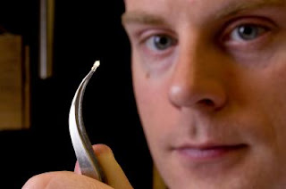
David Rand, Brown University

Caption: David Rand is a professor of biology at Brown University.

Credit: Brown University. Usage Restrictions: None.
Scientists are unsure whether any of these afflictions led directly to the flies' death.

A separate experiment showed adult flies transported carbon nanoparticles and then deposited them elsewhere when they groomed themselves.

The findings, published online in Environmental Science & Technology, help to show the risks of carbon nanoparticles in the environment, said David Rand, professor of biology, who specializes in fruit fly evolution.

"The point is these same compounds that were not toxic to the (fruit fly) larvae were toxic to the adults in some cases, so there may be analogies to other toxic effects from fine particles,"
Rand, a co-corresponding author, said. "It may be like being in a coal mine. You get sick more from the effects of dust particles than from specific toxins in the dust."

The scientists immersed adult Drosophila in a control test tube and test tubes containing four different types of carbon nanoparticles corresponding with their commercial uses — carbon black (a powder much like printer toner), C60 (spherical molecules known as carbon buckyballs, named for Buckminster Fuller's geodesic designs), single-walled carbon nanotubes, and multiwalled carbon nanotubes. Flies in the test tubes with no carbon nanoparticles, C60 and the multiwalled nanotubes climbed up the tubes with few or no difficulties. But the batches of flies immersed in the carbon black and single-walled nanotubes could not escape their surroundings and died within six to 10 hours, the Brown scientists report.
The causes of death are unclear, but detailed analyses led by chemistry graduate student and lead author Xinyuan Liu showed the flies were affected physically. In some, the carbon nanoparticles covered them from wings to legs, which may have impeded their movement or weighted them down too much to climb. In others, the nanoparticles clogged their breathing holes, or spiracles, which may have suffocated them. In other adults, the nanoparticles covered the surface of their compound eyes, which may have blinded them.

The nanoparticles "glom onto the flies," Rand noted while watching a video of flies in the test tubes. "They just can't move. It's like a dinosaur falling into a tar pit."
Nanoparticles in Flies

Caption: While fly larvae appear to have ingested carbon nanostructures without harm, the nanostructures remained in their bodies through adulthood, raising questions about accumulation in the food chain.

Credit: David Rand laboratory, Brown University. Usage Restrictions: None.
Rand and Robert Hurt, director of Brown's Institute for Molecular and Nanoscale Innovation and the other corresponding author, said the findings are important, because they show that permutations of the same material — carbon — can have different effects in the environment.
Toxic Flies

Caption: Microscopy shows a clean foot and leg of a fruit fly (left), and a foot and leg covered with carbon nanostructures (arrows). Adhering nanostructures may have impeded movement, respiration and vision in adult flies but did not appear toxic to fly larvae that ingested it.

Credit: David Rand laboratory, Brown University. Usage Restrictions: None.
"It's not the nanoparticle per se (that may be hazardous), but the form the nanoparticle is in," Rand said.

In another experiment led by Daniel Vinson, an undergraduate student in engineering, adult Drosophila coated in multiwalled carbon nanotubes carried the carbon on their bodies from one test tube into another and deposited some of the particles in the clean tube. That test showed how insects could be vectors for transporting nanomaterials, Rand said.
While two generations of fruit fly larvae showed no ill effects from eating carbon nanoparticles, the Brown scientists noticed that some of the particles ended up being stored in the flies' tissue. That means the nanoparticles could accumulate as they are passed up the food chain, Rand said.

The researchers have several related experiments in the works. They plan to test fruit flies' response to nanosilver and other nanomaterials with different chemistries, and they will investigate why the adult Drosophila died from varieties of the carbon nanoparticles. ###

The research was funded by the National Science Foundation through a Nanoscale Interdisciplinary Research Teams (NIRT) grant, the National Institute of Environmental Health Sciences, the Superfund Research Program Grant, and the Research Seed Fund Program of Brown's Office of Vice President for Research. Dawn Abt, a research assistant in Rand's lab, contributed to the paper.

Contact: Richard Lewis Richard_Lewis@Brown.edu 401-863-3766 Brown University

Monday, September 28, 2009

Nanoscale origami from DNA

Researchers develop a new toolbox for nano-engineering.

Scientists at the Technische Universitaet Muenchen (TUM) and Harvard University have thrown the lid off a new toolbox for building nanoscale structures out of DNA, with complex twisting and curving shapes. In the August 7 issue of the journal Science, they report a series of experiments in which they folded DNA, origami-like, into three dimensional objects including a beachball-shaped wireframe capsule just 50 nanometers in diameter.

"Our goal was to find out whether we could program DNA to assemble into shapes that exhibit custom curvature or twist, with features just a few nanometers wide," says biophysicist Hendrik Dietz, a professor at the Technische Universitaet Muenchen. Dietz's collaborators in these experiments were Professor William Shih and Dr. Shawn Douglas of Harvard University. "It worked," he says, "and we can now build a diversity of three-dimensional nanoscale machine parts, such as round gears or curved tubes or capsules. Assembling those parts into bigger, more complex and functional devices should be possible."

Origami-like Nanostructures Made from DNA

Caption: Scientists at the Technische Universitaet Muenchen and Harvard University have thrown the lid off a new toolbox for building nanoscale structures out of DNA, with complex twisting and curving shapes. In the Aug 7 issue of the journal Science, they report a series of experiments in which they folded DNA, origami-like, into 3-D objects including a beach ball-shaped wireframe capsule just 50 nanometers in diameter.

Credit: H. Dietz, TUM Dept. of Physics, all rights reserved. Usage Restrictions: Used by permission of H. Dietz, TUM Dept. of Physics, all rights reserved.
As a medium for nanoscale engineering, DNA has the dual advantages of being a smart material – not only tough and flexible but also programmable – and being very well characterized by decades of study. Basic tools that Dietz, Douglas, and Shih employ are programmable self-assembly – directing DNA strands to form custom-shaped bundles of cross-linked double helices – and targeted insertions or deletions of base pairs that can give such bundles a desired twist or curve. Right-handed or left-handed twisting can be specified. They report achieving precise, quantitative control of these shapes, with a radius of curvature as tight as 6 nanometers.

The toolbox they have developed includes a graphical software program that helps to translate specific design concepts into the DNA programming required to realize them. Three-dimensional shapes are produced by "tuning" the number, arrangement, and lengths of helices.
In their current paper, the researchers present a wide variety of nanoscale structures and describe in detail how they designed, formed, and verified them. "Many advanced macroscopic machines require curiously shaped parts in order to function," Dietz says, "and we have the tools to make them. But we currently cannot build something intricate such as an ant's leg or, much smaller, a ten-nanometer-small chemical plant such as a protein enzyme. We expect many benefits if only we could build super-miniaturized devices on the nanoscale using materials that work robustly in the cells of our bodies – biomolecules such as DNA." ###

Original paper: "Folding DNA into Twisted and Curved Nanoscale Shapes," by Hendrik Dietz, Shawn M. Douglas, and William M. Shih, published in the August 7, 2009, issue of Science.

Contact: Prof. Hendrik Dietz. Department of Physics. Technische Universitaet Muenchen
James-Franck-Str. 1. 85748 Garching, Germany. Tel. +49 89 289 12539. Fax: +49 89 289 12523 E-mail: dietz@ph.tum.de www: bionano.physik.tu-muenchen.de

Contact: Patrick Regan regan@zv.tum.de 49-892-892-2743 Technische Universitaet Muenchen

Sunday, September 27, 2009

Plastics that convert light to electricity could have a big impact

Researchers the world over are striving to develop organic solar cells that can be produced easily and inexpensively as thin films that could be widely used to generate electricity.

But a major obstacle is coaxing these carbon-based materials to reliably form the proper structure at the nanoscale (tinier than 2-millionths of an inch) to be highly efficient in converting light to electricity. The goal is to develop cells made from low-cost plastics that will transform at least 10 percent of the sunlight that they absorb into usable electricity and can be easily manufactured.

A research team headed by David Ginger, a University of Washington associate professor of chemistry, has found a way to make images of tiny bubbles and channels, roughly 10,000 times smaller than a human hair, inside plastic solar cells. These bubbles and channels form within the polymers as they are being created in a baking process, called annealing, that is used to improve the materials' performance.

David Ginger, University of Washington

Caption: David Ginger, a University of Washington associate professor of chemistry, displays the tiny probe for a conductive atomic force microscope, used to record photocurrents on scales of millionths of an inch in carbon-based solar cells.

Credit: Mary Levin/University of Washington. Usage Restrictions: News use only.
The researchers are able to measure directly how much current each tiny bubble and channel carries, thus developing an understanding of exactly how a solar cell converts light into electricity. Ginger believes that will lead to a better understanding of which materials created under which conditions are most likely to meet the 10 percent efficiency goal.

As researchers approach that threshold, nanostructured plastic solar cells could be put into use on a broad scale, he said. As a start, they could be incorporated into purses or backpacks to charge cellular phones or mp3 players, but eventually they could make in important contribution to the electrical power supply.
Most researchers make plastic solar cells by blending two materials together in a thin film, then baking them to improve their performance. In the process, bubbles and channels form much as they would in a cake batter. The bubbles and channels affect how well the cell converts light into electricity and how much of the electric current actually gets to the wires leading out of the cell. The number of bubbles and channels and their configuration can be altered by how much heat is applied and for how long.

The exact structure of the bubbles and channels is critical to the solar cell's performance, but the relationship between baking time, bubble size, channel connectivity and efficiency has been difficult to understand. Some models used to guide development of plastic solar cells even ignore the structure issues and assume that blending the two materials into a film for solar cells will produce a smooth and uniform substance. That assumption can make it difficult to understand just how much efficiency can be engineered into a polymer, Ginger said.

For the current research, the scientists worked with a blend of polythiophene and fullerene, model materials considered basic to organic solar cell research because their response to forces such as heating can be readily extrapolated to other materials. The materials were baked together at different temperatures and for different lengths of time.

Ginger is the lead author of a paper documenting the work, published online last month by the American Chemical Society journal Nano Letters and scheduled for a future print edition. Coauthors are Liam Pingree and Obadiah Reid of the UW. The research was funded by the National Science Foundation and the U.S. Department of Energy.

Ginger noted that the polymer tested is not likely to reach the 10 percent efficiency threshold. But the results, he said, will be a useful guide to show which new combinations of materials and at what baking time and temperature could form bubbles and channels in a way that the resulting polymer might meet the standard.

Such testing can be accomplished using a very small tool called an atomic force microscope, which uses a needle similar to the one that plays records on an old-style phonograph to make a nanoscale image of the solar cell. The microscope, developed in Ginger's lab to record photocurrent, comes to a point just 10 to 20 nanometers across (a human hair is about 60,000 nanometers wide). The tip is coated with platinum or gold to conduct electrical current, and it traces back and forth across the solar cell to record the properties.

As the microscope traces back and forth over a solar cell, it records the channels and bubbles that were created as the material was formed. Using the microscope in conjunction with the knowledge gained from the current research, Ginger said, can help scientists determine quickly whether polymers they are working with are ever likely to reach the 10 percent efficiency threshold.

Making solar cells more efficient is crucial to making them cost effective, he said. And if costs can be brought low enough, solar cells could offset the need for more coal-generated electricity in years to come.

"The solution to the energy problem is going to be a mix, but in the long term solar power is going to be the biggest part of that mix," he said. ###

For more information, contact Ginger at 206-685-2331 or ginger@chem.washington.edu.

Contact: Vince Stricherz vinces@u.washington.edu 206-543-2580 University of Washington

Friday, September 25, 2009

Nanoparticles cross blood-brain barrier to enable 'brain tumor painting'

Brain cancer is among the deadliest of cancers. It's also one of the hardest to treat. Imaging results are often imprecise because brain cancers are extremely invasive. Surgeons must saw through the skull and safely remove as much of the tumor as they can. Then doctors use radiation or chemotherapy to destroy cancerous cells in the surrounding tissue.

Researchers at the University of Washington have been able to illuminate brain tumors by injecting fluorescent nanoparticles into the bloodstream that safely cross the blood-brain barrier – an almost impenetrable barrier that protects the brain from infection. The nanoparticles remained in mouse tumors for up to five days and did not show any evidence of damaging the blood-brain barrier, according to results published this week in the journal Cancer Research.

Brain Imaging

Caption: This image shows a mouse brain tumor imaged using nanoparticles (left column) or conventional techniques (right column) combined with optical imaging and MRI. The nanoparticles give a clearer picture of the tumor, which is located at the back of the brain in the cerebellum.

Credit: University of Washington. Usage Restrictions: With credit to University of Washington.
Results showed the nanoparticles improved the contrast in both MRI and optical imaging, which is used during surgery.

"Brain cancers are very invasive, different from the other cancers. They will invade the surrounding tissue and there is no clear boundary between the tumor tissue and the normal brain tissue," said lead author Miqin Zhang, a UW professor of materials science and engineering.

Being unable to distinguish a boundary complicates the surgery. Severe cognitive problems are a common side effect.

"If we can inject these nanoparticles with infrared dye, they will increase the contrast between the tumor tissue and the normal tissue," Zhang said. "So during the surgery, the surgeons can see the boundary more precisely.
"We call it 'brain tumor illumination or brain tumor painting,'" she said. "The tumor will light up."

Nano-imaging could also help with early cancer detection, Zhang said. Current imaging techniques have a maximum resolution of 1 millimeter (1/25 of an inch). Nanoparticles could improve the resolution by a factor of 10 or more, allowing detection of smaller tumors and earlier treatment.

Until now, no nanoparticle used for imaging has been able to cross the blood-brain barrier and specifically bind to brain-tumor cells. With current techniques doctors inject dyes into the body and use drugs to temporarily open the blood-brain barrier, risking infection of the brain.

The UW team surmounted this challenge by building a nanoparticle that remains small in wet conditions. The particle was about 33 nanometers in diameter when wet, about a third the size of similar particles used in other parts of the body.

Crossing the blood-brain barrier depends on the size of the particle, its lipid, or fat, content, and the electric charge on the particle. Zhang and colleagues built a particle that can pass through the barrier and reach tumors. To specifically target tumor cells they used chlorotoxin, a small peptide isolated from scorpion venom that many groups, including Zhang's, are exploring for its tumor-targeting abilities. On the nanoparticle's surface Zhang placed a small fluorescent molecule for optical imaging, and binding sites that could be used for attaching other molecules.

Future research will evaluate this nanoparticle's potential for treating tumors, Zhang said. She and colleagues already showed that chlorotoxin combined with nanoparticles dramatically slows tumors' spread. They will see whether that ability could extend to brain cancer, the most common solid tumor to affect children.

Merely improving imaging, however, would improve patient outcomes.

"Precise imaging of brain tumors is phenomenally important. We know that patient survival for brain tumors is directly related to the amount of tumor that you can resect," said co-author Richard Ellenbogen, professor and chair of neurological surgery at the UW School of Medicine. "This is the next generation of cancer imaging," he said. "The last generation was CT, this generation was MRI, and this is the next generation of advances." ###

Other co-authors are Omid Veiseh, Conroy Sun, Chen Fang, Narayan Bhattarai, Jonathan Gunn of the UW's department of materials science and engineering; Forrest Kievit and Kim Du of UW bioengineering; Donghoon Lee of UW radiology; Barbara Pullar of the Fred Hutchinson Cancer Research Center; and Jim Olson of the Fred Hutchinson Cancer Research Center and Seattle Children's Hospital.

The research was funded by the National Institutes of Health, the Jordyn Dukelow Memorial Fund and the Seattle Children's Hospital Brain Tumor Research Endowment.

Contact: Hannah Hickey hickeyh@uw.edu 206-543-2580 University of Washington

Thursday, September 24, 2009

Rapid heating prepares energy-saving zeolite for greater role in industrial separations

New technique eliminates grain boundary defects, researchers report in Science

Thin-film zeolite membranes with tiny, molecule-sized pores are one step closer to replacing the energy-intensive processes now used in industrial separations, a group of academic researchers is reporting.

Writing this week in Science magazine, the group says the membranes' ability to separate molecules in a mixture is significantly improved by subjecting the zeolite to rapid thermal processing (RTP). By heating the membranes from room temperature to 700 degrees Centigrade in one minute, maintaining this temperature for up to two minutes and then quickly cooling it, the researchers say they have been able to eliminate the formation of grain boundary defects that undermine the sieve-like quality of zeolite's uniformly sized nanopores.

Michael Tsapatsis

Michael Tsapatsis, Professor, Chemical Engineering and Materials Science
Office: 445 Amundson Hall. Phone: (612) 626-0920, Email: tsapatsis@cems.umn.edu
The research group, led by Michael Tsapatsis, Amundson Chair Professor of chemical engineering and materials science at the University of Minnesota, says RTP shows promise in achieving greater yield and energy efficiency in zeolite membrane production.

Tsapatsis' group reported its results in an article titled "Grain Boundary Defect Elimination in a Zeolite Membrane by Rapid Thermal Processing." The article was coauthored by Tsapatsis; Jungkyu Choi, formerly of the University of Minnesota and now a postdoctoral fellow at the University of California at Berkeley; Hae-Kwon Jeong, assistant professor of chemical engineering at Texas A&M University;
Mark A. Snyder, assistant professor of chemical engineering at Lehigh University in Bethlehem, Pennsylvania; Jared A. Stoeger, a graduate student at the University of Minnesota; and Richard I. Masel, professor of chemical and biomolecular engineering at the University of Illinois at Urbana-Champaign.

Zeolites are crystalline aluminosilicate materials whose compositions and nanoporous structures can be fine-tuned for applications in catalysis, adsorption and ion exchange. They are called "molecular sieves" because their pores, being small and very uniform in size, can sort and separate molecules selectively according to the molecules' size.

As microscopic particles, zeolites are used in a variety of applications, including the creation of pure streams of oxygen and other gases, the catalytic cracking of petroleum into gasoline, water purification and softening, the dewatering of ethanol, and as additives in laundry detergents.

Zeolite membranes are commonly formed by depositing zeolite crystals on a porous surface and inter-growing these crystals into a continuous film. Several challenges have kept zeolite membranes from achieving their full industrial potential. These include high processing costs; scalability, or the ability to make zeolite membranes in large area; and the difficulty in controlling grain boundary defects, or non-selective pathways at the crystal grain interfaces, which cause poor separation performance.

Meanwhile, an estimated 15 percent of the world's energy consumption is used for the industrial separations of molecules and mixtures, often in volatile, energy-hungry distillation towers. By contrast, zeolite membranes with optimal porosity consume much less energy when they perform separations.

When zeolites are made, structure directing agents, or SDAs, direct the formation of the porous crystalline structure. But the SDAs are then trapped inside the zeolite pores in what scientists call a "ship in the bottle" effect. These SDAs block the zeolite pores and must be removed so other molecules can pass through. High-temperature treatment is typically used to remove the pores, but the heat has little effect on the zeolite, which is stable. But the SDAs, being organic, break up and are removed during heating.

This heat processing must be carried out after the formation of zeolite membranes. Scientists have long believed that this must be done slowly to prevent cracks and other grain boundary defects from forming in the thin film. But the gradual heating promotes the formation of flexible grain boundary defects, or interfaces, between zeolite crystals. These defects can grow much larger than the zeolite pores to become "nonselective" pathways that can be permeated by the very molecules the zeolite is designed to separate.

"The molecules that you are trying to separate with your zeolite film," says Snyder, "can now circumvent the highly selective pathways and pass through the grain boundaries. This has been a major issue for the commercial viability of zeolite membranes."

Tsapatsis' group uses a regimen of RTP to remove the SDA molecules from inside the zeolite pores and to promote what they believe could be chemical "gluing" of the zeolite crystal domains. After heating the zeolite to 700 degrees C, the researchers hold it at that temperature for up to 2 minutes and then cool the material rapidly to room temperature.

The researchers believe the rapid rise in temperature may cause bonding between the crystal domains. "This could possibly decrease the flexibility of the grain boundaries so that they no longer open up between the crystals during operation of the membrane," says Snyder, who worked with Tsapatsis as a postdoctoral researcher. "In short, this type of heat treatment may chemically glue the crystal domains together."

A second round of RTP or conventional slow-heating is then necessary, says Snyder, to completely remove the SDA molecules from inside the pores.

Snyder has used an optical microscopy technique called laser scanning confocal microscopy to examine the grain boundaries in conventionally produced zeolite membranes and in zeolite membranes that have undergone RTP. Confocal microscopy is used widely in biology research to take optical slices of transparent, 3-D structures. Snyder and Tsapatsis were among the first teams to use the technique to characterize zeolite membranes by selectively labeling grain boundary defects with fluorescing molecules. The zeolite they study is silicate-1, one of the more than 170 frameworks of zeolite, which has pores measuring .56 nm in diameter.

"Confocal microscopy shows us the nonselective pathways and other defect features within the membrane," says Snyder.

Confocal microscopy enabled Tsapatsis' group to understand why RTP-treated zeolite membranes achieved a better separation performance than conventionally processed membranes. While a high density of grain boundary defects were observed in conventionally treated membranes, very few defect features were identified in the RTP-treated films. This suggests that grain boundaries in the RTP-treated films are either smaller or less flexible.

This apparent decrease in the number, size and flexibility of grain boundaries in the RTP-treated membranes influences the achievable resolution of the molecular separations, the researchers say.

"A challenging separation that is commonly used to test zeolite membranes," says Snyder, "is that of the isomers orthoxylene and paraxylene. "Orthoxylene is slightly larger and should not pass readily through the pores. One would expect a perfect zeolite membrane to allow a permeation rate for paraxylene that is about two orders of magnitude higher than that of orthoxylene."

"A considerable increase in paraxylene/orthoxylene selectivity was observed for RTP-treated membranes," the researchers wrote in Science, "resulting in an attractive combination of paraxylene permeance and mixture separation factor."

The researchers concluded their article by saying, "If its beneficial effects on performance can be demonstrated for other zeolite types, compositions and microstructures, RTP could contribute, in combination with fast one-step deposition methods, to the realization of large-scale production of zeolite membranes." ###

Contact: Kurt Pfitzer kap4@lehigh.edu 610-758-3017 Lehigh University

Tuesday, September 22, 2009

Jet-propelled imaging for an ultrafast light source

John Spence, a physicist at Arizona State University, is a longtime user of the Advanced Light Source at Lawrence Berkeley National Laboratory, where he has contributed to major advances in lensless imaging. It's a particularly apt propensity for someone who works with x-rays, since they can't be focused with ordinary lenses.

As new light sources evolve to produce brighter x-rays in faster pulses, lensless imaging becomes ever more critical for science. Among the promises of superbright, ultrafast x-ray pulses is the ability to solve the structure of the complicated molecules from which our bodies are made. All living things are made of proteins and nucleic acids, but relatively few of the atomic structures of the thousands, perhaps millions, of varieties of proteins are known.

Sample Jet Nozzle

Caption: In the gas dynamic virtual nozzle, liquid flows through a internal capillary, issuing some distance from the opening in the outer tube through which the gas flows. Approaching the narrow opening, gas pressure and speed increase, focusing the thin stream of liquid until it is so small only a single protein or virus can fit into each droplet.

Credit: courtesy John Spence, ASU. Usage Restrictions: None.
The Linac Coherent Light Source (LCLS) will soon begin operation at the SLAC National Accelerator Laboratory in Palo Alto, California, using energetic electrons from a linear accelerator to produce coherent x-rays with an instrument called a free electron laser (FEL). The x-rays will be delivered 120 times a second in pulses only a tenth of a trillionth of a second long – about the time it takes light to travel the width of a human hair. These brief, bright pulses offer a novel approach to the problem of protein structure.

Unfolding the origami

Proteins begin as strings of amino acids that fold themselves into an amazing variety of origami-like structures, whose bumps and crannies and distribution of electrical charges determine how they act individually or fit together to form complex molecular machines. Simple organisms like viruses often consist of a few proteins fitted together to enclose a thread of DNA or RNA.

Proteins are usually large molecules containing many thousands of atoms. Drug molecules are much smaller, and do their work by attaching themselves to the larger protein molecules.
A knowledge of the arrangement of a protein's atoms is therefore a great help to drug designers, who like to understand how a drug molecule will dock with a protein to promote or inhibit its activity, or cripple the organism of which it is a part.

Until now, the best way to solve the structure of a protein or virus has been with x-ray crystallography. The crystal consists of many copies of the protein or virus arranged in regular order. As the crystal rotates in the x-ray beam, x-rays scatter off the atoms and reveal – once these complex diffraction patterns have been converted into a 3-D image by computers – how the electrons, and thus the atoms, are arranged.
But many proteins can't be crystallized at all, and others are so difficult to crystallize it's virtually impossible to obtain crystals large enough to use in today's light sources.

Ultrafast, ultrabright x-rays offer a way past this dilemma. The idea is that a quick pulse of tightly focused x-rays can be diffracted from a microcrystal or even a single protein or virus in solution.
Sample Jet Stream

Caption: The frequency at which droplets emerge from the sample jet is controlled by an acoustic trigger, which can be tuned so that each droplet containing a protein or virus meets an incoming pulse of x-rays.

Credit: courtesy John Spence, ASU. Usage Restrictions: None.
The pulse is so brief that it comes and goes before any of the atoms can move, freezing their orientation like a strobe light. Just as important, a sufficiently brief pulse may terminate before radiation damage effects can start. In this way it can outrun radiation damage, always one of the fundamental limitations to imaging in biology.

Another quick pulse could be diffracted from another copy of the protein in a different orientation. As the process is repeated, diffractions from different angles give the overlapping views needed for the computer to construct a 3-D image of the structure.

It's a great idea, but as Spence notes, there are a few problems. "So as not to scatter, the x-ray beam has to be in a high vacuum, but a protein or virus in its natural state is usually wet. As in T. S. Eliot's Wasteland, water is life. How do we maintain the protein or virus in an aqueous environment inside the vacuum?"

Shot from a microcannon

The answer was what Spence calls a "particle gun, like an ink-jet printer," designed to inject a beam of water droplets across the tightly focused x-ray beam in single file, each droplet so small it contains only a single protein or virus. He and colleagues Bruce Doak and Uwe Weierstall of ASU designed a nozzle that can fire liquid droplets, each less than a millionth of a meter in diameter (one micrometer), faster than hundreds of thousands of times a second. The sample jet is designed to shoot droplets right through a pulsed beam of x-rays a billion times brighter than any ever created in a light source before.

ASU postdoc Dan DePonte did most of the hard work and refinement needed to realize the design. It wasn't easy. Nozzles made of solid material like glass invariably clog up, limiting droplets to at best 20 micrometers across. What Spence and his colleagues wanted was a jet of particles less than a micrometer in size.

Back in 1878 Lord Rayleigh, a professor of experimental physics at Cambridge University, discovered that a smooth, cylindrical jet of liquid emerging from an orifice spontaneously breaks up to form a train of spherical droplets. In the late 1990s, physicist Alfonso Gañán-Calvo of the University of Seville found a way to surround the streaming liquid with pressurized gas to make a co-flowing liquid sheath. By adjusting gas and liquid pressure and other parameters, he was able to create a "virtual nozzle" that could shrink the diameter of the liquid jet to a thread so small it would not clog the physical aperture of the tube. In effect, the gas sheath acts to focus the liquid stream.

Spence and his colleagues needed a true microthread of liquid, however, one that produced droplets sized a millionth of a meter or less. In their nozzle, liquid flows through a narrow capillary inside the tube through which the gas flows; the liquid issues from the capillary some distance from the opening in the outer tube, so the gas surrounds it, then increases speed and pressure as it approaches the opening, squeezing and accelerating the thin stream of liquid until it is so small that the proteins or viruses dissolved in the liquid can only fit into the droplets one at a time.

And the nozzle won't clog, because even a particle bigger than the sample protein or virus – bigger than the stream of liquid itself – can still fly through the glass nozzle without hitting the walls and getting stuck.

The frequency at which the droplets emerge can be controlled by an oscillator the researchers call an "acoustic trigger." Tuning the acoustic trigger adjusts the frequency so that each droplet containing a protein or virus meets an incoming pulse of x-rays.

The entire device – which the researchers call a gas dynamic virtual nozzle (GDVN) – is only about a millimeter in diameter (not counting feed lines and cables) and fits to the side of the beamline's vacuum chamber. After passing through the beam, the liquid droplets and the gas (typically carbon dioxide) freeze in a trap opposite the injection point, without significantly reducing the vacuum.

In 2008 Spence and his colleagues, including Berkeley Lab's David Shapiro, successfully tested the GDVN on the Advanced Light Source beamline 9.0.1, managed by Berkeley Lab's Stefano Marchesini. The test was done with protein microcrystals extracted from the fluid in which researchers were attempting to grow larger crystals. These are the smallest protein nanocrystals from which diffraction patterns have ever been obtained, and the first from membrane protein nanocrystals – among the most resistant to crystallization.

Although the microcrystals weren't individual protein specimens, and while the 9.0.1's x-ray beams aren't as bright or as rapidly pulsed as SLAC's LCLS will be, the experiment demonstrated the jet technique's high potential for speeds and exposures that won't subject the samples to radiation damage. Some of the patterns the researchers obtained come from nanocrystals just a few molecules on a side, with a width of about 100 billionths of a meter (100 nanometers). At SLAC, the researchers plan to steadily reduce the nanocrystal size down to single molecules.

The corresponding reduction in scattered intensity will hasten and improve lensless imaging. The first step in lensless imaging is scattering the beam from the sample; the second step is constructing the image by interpreting and combining the data from the diffracted x-rays.

In order to merge the different views (projections) of an object, which is subsequently vaporized in this "diffract-and-destroy" mode, it is important that they all be identical. In biology, that leaves only molecules like proteins and viruses. DNA or RNA inside a virus is often packed differently in each virus, and cells are not identical at the molecular level, so cannot be studied in 3-D by this method.

Besides identical particles, successful data-merging also depends partly on knowing how the sample was oriented in the beam – easy to do with a large crystal, not so easy to do with a sample inside a drop of liquid whizzing across the beam. It may be possible to orient flying droplets by optical methods such as polarized laser beams or with specially shaped nozzles.

Perhaps simpler is to use the ever-increasing power of the computer – which for a lensless imaging system is where most of the functions of a lens reside. Computer systems have been developed that infer the orientation of the sample from the diffraction pattern itself, even when as few as four percent of the pixels in the detector light up. It does take a lot of diffraction patterns to derive an image this way – as many as 10 million – which will take the LCLS a few hours until better ways of orienting the droplets can be advised.

Nevertheless, Spence's group recently obtained excellent diffraction patterns of MS2 virus capsids at the ALS by subtracting the diffraction "noise" of the liquid jet itself. These capsids are the shells of the virus lacking its RNA genome and have the regular shape of buckyballs. Eventually the LCLS will be able to get a good diffraction pattern from a target like this with a single ultrabright pulse. In this case, however, computer processing was able to derive an excellent pattern by averaging diffraction from a series of samples.

DePonte will soon install Spence and Doak's ultrafine, ultrafast "inkjet printer," tested at the ALS, on the powerful new SLAC machine. It will be the first step into a bold new future for investigating the biological universe, one big molecule at a time. ###

Berkeley Lab is a U.S. Department of Energy national laboratory located in Berkeley, California. It conducts unclassified scientific research for DOE's Office of Science and is managed by the University of California. Visit our website at www.lbl.gov.

Contact: Paul Preuss paul_preuss@lbl.gov 510-486-6249 DOE/Lawrence Berkeley National Laboratory

Sunday, September 20, 2009

All-in-1 nanoparticle: A Swiss Army knife for nanomedicine

Nanoparticles are being developed to perform a wide range of medical uses – imaging tumors, carrying drugs, delivering pulses of heat. Rather than settling for just one of these, researchers at the University of Washington have combined two nanoparticles in one tiny package.

The result is the first structure that creates a multipurpose nanotechnology tool for medical imaging and therapy. The structure is described in a paper published online this week in the journal Nature Nanotechnology.

"This is the first time that a semiconductor and metal nanoparticles have been combined in a way that preserves the function of each individual component," said lead author Xiaohu Gao, a UW assistant professor of bioengineering.

Multifunctional Nanoparticle

Caption: This is a quantum dot encapsulated in a gold shell. The total structure measures less than 20 nanometers across.

Credit: University of Washington, Usage Restrictions: None.
The current focus is on medical applications, but the researchers said multifunctional nanoparticles could also be used in energy research, for example in solar cells.

Quantum dots are fluorescent balls of semiconductor material just a few nanometers across, a small fraction of the wavelength of visible light (a nanometer is 1-millionth of a millimeter). At this tiny scale, quantum dots' unique optical properties cause them to emit light of different colors depending on their size. The dots are being developed for medical imaging, solar cells and light-emitting diodes.
Glowing gold nanoparticles have been used since ancient times in stained glass; more recently they are being developed for delivering drugs, for treating arthritis and for a type of medical imaging that uses infrared light. Gold also reradiates infrared heat and so could be used in medical therapies to cook nearby cells.

But combine a quantum dot and a gold nanoparticle, and the effects disappear. The electrical fields of the particles interfere with one another and so neither behaves as it would on its own. The two have been successfully combined on a surface, but never in a single particle.

The paper describes a manufacturing technique that uses proteins to surround a quantum dot core with a thin gold shell held at 3 nanometers distance, so the two components' optical and electrical fields do not interfere with one another. The quantum dot likely would be used for fluorescent imaging. The gold sphere could be used for scattering-based imaging, which works better than fluorescence in some situations, as well as for delivering heat therapy.

The manufacturing technique developed by Gao and co-author Yongdong Jin, a UW postdoctoral researcher, is general and could apply to other nanoparticle combinations, they said.

"We picked a tough case," Gao said. "It is widely known that gold or any other metal will quench quantum dot fluorescence, eliminating the quantum dot's purpose."

Gao and Jin avoided this problem by building a thin gold sphere that surrounds but never touches the quantum dot. They carefully controlled the separation between the gold shell and the nanoparticle core by using chains of polymer, polyethylene glycol. The distance between the quantum dot core and charged gold ion is determined by the length of the polymer chain and can be increased with nanometer precision by adding links to the chain. On the outside layer they added short amino acids called polyhistidines, which bind to charged gold atoms.

Gao compares the completed structure to a golden egg, where the quantum dot is the yolk, the gold is the shell, and polymers fill up the space of the egg white.

Using ions allowed the researchers to build a 2- to 3-nanometer gold shell that's thin enough to allow about half of the quantum dot's fluorescence to pass through.

"All the traditional techniques use premade gold nanoparticles instead of gold ions," Gao said. "Gold nanoparticles are 3 to 5 nanometers in diameter, and with factoring in roughness the thinnest coating you can build is 5-6 nanometers. Gold ions are much, much smaller."

The total diameter of the combined particle is roughly 15-20 nanometers, small enough to be able to slip into a cell.

Incorporating gold provides a well-established binding site to attach biological molecules that target particular cells, such as tumor cells. Gold could also potentially amplify the quantum dot's fluorescence by five to 10 times, as it has in other cases.

The gold sphere offers one further benefit. Gold is biocompatible, is medically approved and does not biodegrade. A gold shell could thus provide a durable non-toxic container for nanoparticles being used in the body, Gao said. ###

The research was supported by the National Institutes of Health, the National Science Foundation, the Seattle Foundation and the UW's Department of Bioengineering.

For more information, contact Gao at 206-543-6562 or xgao@u.washington.edu.

Contact: Hannah Hickey hickeyh@uw.edu 206-543-2580 University of Washington

Saturday, September 19, 2009

Nanodiamonds deliver insulin for wound healing

Bacterial infection is a major health threat to patients with severe burns and other kinds of serious wounds such as traumatic bone fractures. Recent studies have identified an important new weapon for fighting infection and healing wounds: insulin.

Now, using tiny nanodiamonds, researchers at Northwestern University have demonstrated an innovative method for delivering and releasing the curative hormone at a specific location over a period of time. The nanodiamond-insulin clusters hold promise for wound-healing applications and could be integrated into gels, ointments, bandages or suture materials.

Dean Ho

“This study introduces the concept of nanodiamond-mediated release of therapeutic proteins,” said Dean Ho (right).
Localized release of a therapeutic is a major challenge in biomedicine. The Northwestern method takes advantage of a condition typically found at a wound site -- skin pH levels can reach very basic levels during the repair and healing process. The researchers found that the insulin, bound firmly to the tiny carbon-based nanodiamonds, is released when it encounters basic pH levels, similar to those commonly observed in bacterially infected wounds.
These basic pH levels are significantly greater than the physiological pH level of 7.4.

The results of the study were published online July 26 by the journal Biomaterials.

"This study introduces the concept of nanodiamond-mediated release of therapeutic proteins," said Dean Ho, assistant professor of biomedical engineering and mechanical engineering at the McCormick School of Engineering and Applied Science. Ho led the research. "It's a tricky problem because proteins, even small ones like insulin, bind so well to the nanodiamonds. But, in this case, the right pH level effectively triggers the release of the insulin."

A substantial amount of insulin can be loaded onto the nanodiamonds, which have a high surface area. The nanodiamond-insulin clusters, by releasing insulin in alkaline wound areas, could accelerate the healing process and decrease the incidence of infection. Ho says this ability to release therapeutics from the nanodiamonds on demand represents an exciting strategy towards enhancing the specificity of wound treatment.

In their studies, Ho and his colleagues showed that the insulin was very tightly bound to the nanodiamonds when in an aqueous solution near the normal physiological pH level. Measurements of insulin function revealed that the protein was virtually inactive when bound to the nanodiamonds -- a beneficial property for preventing excess or unnecessary drug release.

Upon increasing the pH to the basic levels commonly observed in the skin during severe burns, the researchers confirmed the insulin was released from the nanodiamond clusters and retained its function. Exploiting this pH-mediated release mechanism may provide unique advantages for enhanced drug delivery methods.

The researchers also found the insulin slowly and consistently released from the nanodiamond clusters over a period of several days.

Insulin accelerates wound healing by acting as a growth hormone. It encourages skin cells to proliferate and divide, restores blood flow to the wound, suppresses inflammation and fights infection. Earlier investigations have confirmed an increase in alkalinity of wound tissue, due to bacterial colonization, to levels as high as pH 10.5, the pH level that promoted insulin release from the nanodiamonds in the Northwestern study.

Ho's group next will work on integrating the nanodiamond-insulin complexes into a gel and conducting preclinical studies. The researchers also will investigate different areas of medicine in which the nanodiamond-insulin clusters could be used.

Nanodiamonds have many advantages for biomedical applications. The large surface area allows a large amount of therapeutic to be loaded onto the particles. They can be functionalized with nearly any type of therapeutic, including small molecules, proteins and antibodies. They can be suspended easily in water, an important property in biomedicine. The nanodiamonds, each being four to six nanometers in diameter, are minimally invasive to cells, biocompatible and do not cause inflammation, a serious complication. And they are very scalable and can be produced in large quantities in uniform sizes.

By harnessing the unique surface properties of the nanodiamonds, Ho and his colleagues have demonstrated that the nanodiamonds serve as platforms that can successfully bind, deliver and release several classes of therapeutics, which could impact a broad range of medical needs.

Ho's research group also has studied nanodiamonds for applications in cancer therapy. They demonstrated that nanodiamonds are capable of releasing the chemotherapy agent Doxorubicin in a sustained and consistent manner. (Ho is a member of the Robert H. Lurie Comprehensive Cancer Center of Northwestern University.)

In addition to using the nanodiamonds in their particle form, Ho's group has developed devices that harness the slow drug-release capabilities of the nanodiamonds. More recently, his team has shown that nanodiamonds are effective in dispersing insoluble drugs in water, boosting their potential for broader applications in medicine. ###

The National Science Foundation, the National Institutes of Health, the Wallace H. Coulter Foundation and the V Foundation for Cancer Research supported the research.

The title of the Biomaterials paper is "Nanodiamond-Insulin Complexes as pH-Dependent Protein Delivery Vehicles." In addition to Ho, other authors of the paper are Rafael Shimkunas (first author), Erik Robinson, Robert Lam, Steven Lu, Xiaoyang Xu, Xueqing Zhang and Houjin Huang, all from Northwestern, and Eiji Osawa, from the NanoCarbon Research Institute at Shinshu University, Nagano, Japan.

Contact: Kyle Delaney k-delaney@northwestern.edu 847-467-4010 Northwestern University

Thursday, September 17, 2009

UH Researchers Pressing on in Their Mission to Power the Nanodevices of Tomorrow

Team is Revealing Secrets of Electricity-Producing Materials, Much like humans, materials are capable of some pretty remarkable things when they're placed under pressure. In fact, under the right conditions, materials can even produce electricity.

Driven by the vision of our society one day being basically self-propelled, a team of University of Houston scientists has set out to both amplify and provoke that potential in materials known as piezoelectrics, which naturally produce electricity when literally subjected to strain. The goal is to use piezoelectrics to create nanodevices that can power electronics, such as cell phones, MP3 players and even biomedical implants.

Dr. Pradeep Sharma

Dr. Pradeep Sharma, Bill D. Cook Chair Associate Professor of Mechanical Engineering
Dept. of Mechanical Engineering. University of Houston. Engineering Building One
Houston, TX 77204-4006
"Nanodevices using piezoelectric materials will be light, environmentally friendly and draw on inexhaustible energy supplies," says associate professor Pradeep Sharma, one of the creative minds at the Cullen College of Engineering running two projects on piezoelectrics. "Imagine a sensor on the wing of a plane or a satellite. Do we really want to change its battery every time its power source gets exhausted? Hard-to-access devices could be self-powered."

Piezoelectric materials convert mechanical energy into electrical energy, Sharma explains.

"Indeed, gas lighters used in most homes are based on this," he says. "These future piezoelectric nanodevices will also generate an electrical current in response to mechanical stimuli. Then, the energy will be stored in batteries or, even better, in nanocapacitors for use when needed."
Although piezoelectrics have been used for many years, Sharma's team is exploring new possibilities by beefing up the effect in natural piezoelectrics. Doing so requires understanding the phenomenon that spurs piezoelectricity, known as "flexoelectricity."

"Flexoelectricity, at the nanoscale, allows you to coax ordinary material to behave like a piezoelectric one. Perhaps more importantly, this phenomenon exists in materials that are already piezoelectric. You can make the effect even larger," Sharma says.

For example, the piezoelectricity in barium titanate can be increased by 300 percent when the material is reduced to a 2-nanometer-beam and pressure is applied. "Thus, you'll take an ordinary piezoelectric material and really give it some juice," he says.

Sharma underscores the flexoelectric effect is a function of size - and the smaller the better, at least for generating piezoelectric power. Materials with nanoscale features - such as nanoscale thin plates stacked on each other or materials with particles or holes the size of a few nanometers - exhibit a much larger flexoelectric effect, he says.

Ramanan Krishnamoorti, chair of the department of chemical and biomolecular engineering, is working with Sharma to embed classes of nanostructures in polymers to create unusual types of piezoelectrics.

Meanwhile, Sharma and professor Ken White recently reported that the electrical activity caused by flexoelectricity also affects how a material's resiliency. They tested their theory - that the elasticity of a material would be quite altered by flexoelectricity-caused electrical activity - by poking the material with a sophisticated needle.

"We basically predicted that when you poke it, because of this electrical activity, depending upon how big a crater you create, your elastic behavior will change. It's not supposed to. Ordinarily, whether you make a big crater or small crater, if you calculate how stiff it is or soft it is, it'll give you the same answer - a constant," Sharma says.

White and Sharma conducted several experiments on single crystals of materials.

"By monitoring the stiffness of the material as the crater became larger and larger," White says, "we discovered a change in elasticity relative to size, which could only be explained by flexoelectric effect."

Though a fair amount of research on piezoelectrics has been done, White says, the fabrication of piezoelectric nanostructures remains challenging. The amount of power that can be harvested is still too low to actually power wearable devices, he says, unless efficient electric storage solutions, like nanocapacitors, also are conceived.

Sharma says he would like to see wasted energy be harvested from a variety of sources.

"In principle, any human activities - for example, walking, jumping, swimming - will produce a certain amount of energy," he says, and could be made into electricity by piezoelectric nanostructures in shoes or in backpacks.

White says it's a matter of controlling materials' structures to the point at which considerably more power can be harvested from common activities.

"An enormous benefit can be expected - in everything from soldiers in the field, to police on the street, to air and ground vehicles - in the form of locally powered devices," White explains.

Sharma says the environment contains plenty of waste energy that can be harnessed into useful energy to make ours a "self-powered autonomous society."

"Recent technological advances and breakthroughs play an important role toward achieving that goal, but we need to be patient," he says. "Quantum mechanics, the basis of modern electronics, was ‘discovered' in the early 1900s. Think how long it has taken for us to exploit that." ###

About the University of Houston

The University of Houston, Texas' premier metropolitan research and teaching institution, is home to more than 40 research centers and institutes and sponsors more than 300 partnerships with corporate, civic and governmental entities. UH, the most diverse research university in the country, stands at the forefront of education, research and service with more than 36,000 students.

About the Cullen College of Engineering

The Cullen College of Engineering at UH has produced five U.S. astronauts, 10 members of the National Academy of Engineering, and degree programs that have ranked in the top 10 nationally. With more than 2,600 students, the college offers accredited undergraduate and graduate degrees in biomedical, chemical, civil and environmental, electrical and computer, industrial, and mechanical engineering. It also offers specialized programs in aerospace, materials, petroleum engineering and telecommunications.

Contact: Angela Hopp ahopp@uh.edu 713-743-8153 University of Houston

Tuesday, September 15, 2009

Music is the engine of new U-M lab-on-a-chip device VIDEO

ANN ARBOR, Mich.---Music, rather than electromechanical valves, can drive experimental samples through a lab-on-a-chip in a new system developed at the University of Michigan. This development could significantly simplify the process of conducting experiments in microfluidic devices.

A paper on the research was published online in the Proceedings of the National Academy of Sciences the week of July 20.

A lab-on-a-chip, or microfluidic device, integrates multiple laboratory functions onto one chip just millimeters or centimeters in size. The devices allow researchers to experiment on tiny sample sizes, and also to simultaneously perform multiple experiments on the same material. There is hope that they could lead to instant home tests for illnesses, food contaminants and toxic gases, among other advances.

Mark A. Burns, P.E.

Mark A. Burns, P.E. 3074F Dow (734) 764-1516, FAX: (734) 763-0459 maburns@umich.edu, Biochemical separations, field-enhanced separations, microfabricated chemical analysis systems, DNA genotyping and sequencing.
To do an experiment in a microfluidic device today, researchers often use dozens of air hoses, valves and electrical connections between the chip and a computer to move, mix and split pin-prick drops of fluid in the device's microscopic channels and divots.

"You quickly lose the advantage of a small microfluidic system," said Mark Burns, professor and chair of the Department of Chemical Engineering and a professor in the Department of Biomedical Engineering.

"You'd really like to see something the size of an iPhone that you could sneeze onto and it would tell you if you have the flu. What hasn't been developed for such a small system is the pneumatics---the mechanisms for moving chemicals and samples around on the device."

The U-M researchers use sound waves to drive a unique pneumatic system that does not require electromechanical valves.
Instead, musical notes produce the air pressure to control droplets in the device. The U-M system requires only one "off-chip" connection.

"This system is a lot like fiberoptics, or cable television. Nobody's dragging 200 separate wires all over your house to power all those channels," Burns said. "There's one cable signal that gets decoded."
The system developed by Burns, chemical engineering doctoral student Sean Langelier, and their collaborators replaces these air hoses, valves and electrical connections with what are called resonance cavities. The resonance cavities are tubes of specific lengths that amplify particular musical notes.

These cavities are connected on one end to channels in the microfluidic device, and on the other end to a speaker, which is connected to a computer. The computer generates the notes, or chords. The resonance cavities amplify those notes and the sound waves push air through a hole in the resonance cavity to their assigned channel. The air then nudges the droplets in the microfluidic device along.


Fight song moves droplets, This video provided by College of Engineering researchers uses the Michigan fight song to demonstrate how sound waves can be used to move droplets through microfluidic devices.
"Each resonance cavity on the device is designed to amplify a specific tone and turn it into a useful pressure," Langelier said. "If I play one note, one droplet moves. If I play a three-note chord, three move, and so on. And because the cavities don't communicate with each other, I can vary the strength of the individual notes within the chords to move a given drop faster or slower."

Burns describes the set-up as the reverse of a bell choir. Rather than ringing a bell to create sound waves in the air, which are heard as music, this system uses music to create sound waves in the device, which in turn, move the experimental droplets.

"I think this is a very clever system," Burns said. "It's a way to make the connections between the microfluidic world and the real world much simpler."

The new system is still external to the chip, but the researchers are working to make it smaller and incorporate it on a microfluidic device. That would be a step closer to a smartphone-sized home flu test. ###

The paper is called, "Acoustically-driven programmable liquid motion using resonance cavities." Other authors are U-M chemical engineering graduate students Dustin Chang and Ramsey Zeitoun. The research is funded by the National Institutes of Health and the National Science Foundation. The University is pursuing patent protection for the intellectual property, and is seeking commercialization partners.

For more information:

Mark Burns: www.engin.umich.edu/dept/cheme/people/burns
Sean Langelier: www.engin.umich.edu/dept/che/research/burns/Sean

Michigan Engineering:

The University of Michigan College of Engineering is ranked among the top engineering schools in the country. At more than $130 million annually, its engineering research budget is one of largest of any public university. Michigan Engineering is home to 11 academic departments and a National Science Foundation Engineering Research Center. The college plays a leading role in the Michigan Memorial Phoenix Energy Institute and hosts the world class Lurie Nanofabrication Facility. Find out more at www.engin.umich.edu/.

Contact: Nicole Casal Moore ncmoore@umich.edu 734-647-1838 University of Michigan

Sunday, September 13, 2009

Reveal the enemy

Carbon nanotubes and aptamers: New biosensor detects extremely low bacteria concentrations quickly, easily and reliably.

Bacterial diseases are usually detected by first enriching samples, then separating, identifying, and counting the bacteria. This type of procedure usually takes at least two days after arrival of the sample in the laboratory. Tests that work faster, in the field, and without complex sample preparation, whilst being precise and error-free, are thus high on the wish list. A Spanish research team headed by Jordi Riu and F. Xavier Rius at the University Rovira i Virgili in Tarragona has now developed a new technique to make this wish come true. With a novel biosensor, they have been able to detect extremely low concentrations of the typhus-inducing Salmonella typhi.

Aptamer Attached to An Electrode Coated with Carbon Nanotubes

Caption: This graphic shows an aptamer attached to an electrode coated with single-walled carbon nanotubes interacts selectively with bacteria. The resulting electrochemical response is highly accurate and reproducible and starts at ultra-low bacteria concentrations, providing a simple, selective method for pathogen detection.

Credit: (C) Wiley-VCH 2009. Usage Restrictions: Permission to use with appropriate credit and link to Credit: (C) Wiley-VCH 2009
As reported in the journal Angewandte Chemie, their new method is based on electrochemical measurements by means of carbon nanotubes equipped with aptamers as bacteria-specific binding sites. If bacteria bind to the aptamers, the researchers detect a change in electrical voltage.

Aptamers are synthetic, short DNA or RNA strands that can be designed and made to bind a specific target molecule. An aptamer that specifically binds to salmonella has recently been developed. The Spanish researchers chose to use this aptamer for their biosensor.
By means of additional functional groups, they securely anchored the aptamers to carbon nanotubes, which were deposited onto an electrode in an ultrathin layer.

In the absence of salmonella, the aptamers fit closely against the walls of the carbon nanotubes. If the biosensor is put into a salmonella-containing sample, the microbes stick to the aptamers like flies to flypaper. This influences the interaction between the aptamers and the nanotubes, which makes a change in the electrode voltage noticeable within seconds.

Using this biosensor, the researchers were able to detect a bacterial concentration equivalent to one salmonella bacterium in 5 mL of medium. Quantitative measurements were possible down to a concentration of about 1000 salmonella per milliliter. This biosensor is specific: it does not react to bacteria other than Salmonella typhi. "Our new technique makes the detection of micro-organisms as fast and simple as the measurement of pH value," say Riu and Rius. ###

Author: Jordi Riu, Universitat Rovira i Virgili, Tarragona (Spain), mailto: jordi.riu@urv.cat

Title: Immediate Detection of Living Bacteria at Ultralow Concentrations Using a Carbon-Nanotube-Based Potentiometric Aptasensor. Angewandte Chemie International Edition, doi: 10.1002/anie.200902090. Copy free of charge. We would appreciate a transcript of your article or a reference to it.

This release is available in German.

Contact: Jordi Riu: jordi.riu@urv.cat, WEB: Wiley-Blackwell

Friday, September 11, 2009

NYU physicists find way to explore microscopic systems through holographic video

Physicists at New York University have developed a technique to record three-dimensional movies of microscopic systems, such as biological molecules, through holographic video. The work, which is reported in Optics Express, has potential to improve medical diagnostics and drug discovery.

The technique, developed in the laboratory of NYU Physics Professor David Grier, is comprised of two components: making and recording the images of microscopic systems and then analyzing these images.

To generate and record images, the researchers created a holographic microscope, which is based on a conventional light microscope. But instead of relying on an incandescent illuminator, which conventional microscopes employ, the holographic microscope uses a collimated laser beam—a beam consisting of a series of parallel rays of light and similar to a laser pointer.

Microscopic Systems Through Holographic Video

In the microscope, a laser beam illuminates the sample. Light scattered by the sample creates an interference pattern which is magnified and recorded. Then measurements of the particle’s position, size, and refractive index are obtained.
When an object is placed into path of the microscope's beam, the object scatters some of the beam's light into a complex diffraction pattern. The scattered light overlaps with the original beam to create an interference pattern reminiscent of overlapping ripples in a pool of water. The microscope then magnifies the resulting pattern of light and dark and records it with a conventional digital video recorder (DVR). Each snapshot in the resulting video stream is a hologram of the original object. Unlike a conventional photograph, each holographic snapshot stores information about the three-dimensional structure and composition of the object that created the scattered light field.
The recorded holograms appear as a pattern of concentric light and dark rings. This resulting pattern contains a wealth of information about the material that originally scattered the light—where it was and what it was comprised of.

Analyzing the images provided a different set of challenges. To do so, the researchers based their work on a quantitative theory explaining the pattern of light that objects scatter. The theory, Lorenz-Mie theory, maintains that the way light is scattered can reveal the size and composition of the object that is scattering it.

"We use that theory to analyze the hologram of each object in the snapshots of our video recording," explained Grier, who is part of NYU's Center for Soft Matter Research. "Fitting the theory to the hologram of a sphere reveals the three-dimensional position of the sphere's center with remarkable resolution. It allows us to view particles a micrometer in size and with nanometric precision—that is, it captures their traits to within one billionth of a meter."

"That's a tremendous amount of information to obtain about a micrometer-scale object, particularly when you consider that you get all of that information in each snapshot," Grier added. "It exceeds other existing technology in terms of tracking particles and characterizing their make-up—and the holographic microscope can do both simultaneously."

Because the analysis is computationally intensive, the researchers employ the number-crunching power of the graphical processing unit (GPU) used in high-end computer video cards. Originally intended to provide high-resolution video performance for computer games, these cards possess capabilities ideal for the holographic microscope.

The team has already employed the technique for a range of applications, from research in fundamental statistical physics to analyzing the composition of fat droplets in milk.

More broadly, the technique creates a more sophisticated method to aid in medical diagnostics and drug discovery. At its most basic level, research in these areas seeks to understand whether or not certain molecular components, i.e., the building blocks of pharmaceuticals, stick together.

One approach, called a "bead-based assay," creates micrometer-scale beads whose surfaces have active groups that bind to the target molecule. Because of their small size, the challenge for researchers is to determine if these beads actually stick to the target molecules. The way this is traditionally done is to create yet another molecule—or tag—that binds to the target molecule. This tag molecule, time-consuming and costly to produce, is typically identified by making it fluorescent or radioactive.

The holographic imaging technique, with its magnification and recording capabilities, allows researchers to observe molecular-scale binding without a tag, saving both time and money. Requiring just one microscopic bead to detect one type of molecule, holographic video microscopy promises a previously unattainable level of miniaturization for medical diagnostic tests and creates possibilities for running very large numbers of sensitive medical tests in parallel. ###

Contact: James Devitt james.devitt@nyu.edu 212-998-6808 New York University

Wednesday, September 09, 2009

Scientists discover repulsive side to light force

New Haven, Conn.—A team of Yale University researchers has discovered a "repulsive" light force that can be used to control components on silicon microchips, meaning future nanodevices could be controlled by light rather than electricity.

The team previously discovered an "attractive" force of light and showed how it could be manipulated to move components in semiconducting micro- and nano-electrical systems—tiny mechanical switches on a chip. The scientists have now uncovered a complementary repulsive force. Researchers had theorized the existence of both the attractive and repulsive forces since 2005, but the latter had remained unproven until now. The team, led by Hong Tang, assistant professor at Yale's School of Engineering & Applied Science, reports its findings in the July 13 edition of Nature Photonics's advanced online publication.

Nano Wave Guides

Caption: Dr. Tang's team shows how interacting lightwaves can be used to control devices on a silicon chip. Credit: Hong Tang/Yale University. Usage Restrictions: Image may be used with appropriate credit given.
"This completes the picture," Tang said. "We've shown that this is indeed a bipolar light force with both an attractive and repulsive component."

The attractive and repulsive light forces Tang's team discovered are separate from the force created by light's radiation pressure, which pushes against an object as light shines on it. Instead, they push out or pull in sideways from the direction the light travels.
Previously, the engineers used the attractive force they discovered to move components on the silicon chip in one direction, such as pulling on a nanoscale switch to open it, but were unable to push it in the opposite direction.

Using both forces means they can now have complete control and can manipulate components in both directions. "We've demonstrated that these are tunable forces we can engineer," Tang said.

In order to create the repulsive force, or the "push," on a silicon chip, the team split a beam of infrared light into two separate beams and forced each one to travel a different length of silicon nanowire, called a waveguide. As a result, the two light beams became out of phase with one another, creating a repulsive force with an intensity that can be controlled—the more out of phase the two light beams, the stronger the force.

"We can control how the light beams interact," said Mo Li, a postdoctoral associate in electrical engineering at Yale and lead author of the paper. "This is not possible in free space—it is only possible when light is confined in the nanoscale waveguides that are placed so close to each other on the chip."

"The light force is intriguing because it works in the opposite way as charged objects," said Wolfram Pernice, another postdoctoral fellow in Tang's group. "Opposite charges attract each other, whereas out-of-phase light beams repel each other in this case."

These light forces may one day control telecommunications devices that would require far less power but would be much faster than today's conventional counterparts, Tang said. An added benefit of using light rather than electricity is that it can be routed through a circuit with almost no interference in signal, and it eliminates the need to lay down large numbers of electrical wires. ###

Funding for the project includes a seed grant from the U.S. Defense Advanced Research Projects Agency and a Young Faculty Award from the National Science Foundation.

Citation: DOI: 10.1038/NPHOTON.2009.116

Contact: Suzanne Taylor Muzzin suzanne.taylormuzzin@yale.edu 203-432-8555 Yale University

Monday, September 07, 2009

Material world: Graphene's versatility promises new applications

Since its discovery just a few years ago, graphene has climbed to the top of the heap of new super-materials poised to transform the electronics and nanotechnology landscape. As N.J. Tao, a researcher at the Biodesign Institute of Arizona State University explains, this two dimensional honeycomb structure of carbon atoms is exceptionally strong and versatile. Its unusual properties make it ideal for applications that are pushing the existing limits of microchips, chemical sensing instruments, biosensors, ultracapacitance devices, flexible displays and other innovations.

In the latest issue of Nature Nanotechnology Letters, Tao describes the first direct measurement of a fundamental property of graphene, known as quantum capacitance, using an electrochemical gate method. A better understanding of this crucial variable should prove invaluable to other investigators participating in what amounts to a gold rush of graphene research.

N. J. Tao, Arizona State University

Caption: N. J. Tao, director of the Center for Bioelectronics and Biosensors at the Biodesign Institute of Arizona State University, has experimentally measured an important property of graphene -- a two-dimensional crystal lattice with broad potential for electronic applications.

Credit: The Biodesign Institute at Arizona State University. Usage Restrictions: None.

two plots of quantum capacitance as a function of potential.

Caption: These are two plots of quantum capacitance as a function of potential. In a) various theoretical predictions of quantum capacitance are shown for graphene samples from pure, (the bottom V-shaped plot) to those above, indicating successively greater levels of impurity. In b) the red curve indicates theoretical prediction of quantum capacitance in graphene as a function of potential. The blue line shows the closely matching results of experimental measurements. Variations from theoretical ideal are the result of charged impurities in the graphene sample.

Credit: The Biodesign Institute at Arizona State University. Usage Restrictions: None.
Although theoretical work on single atomic layer graphene-like structures has been going on for decades, the discovery of real graphene came as a shock. "When they found it was a stable material at room temperature," Tao says, "everyone was surprised." As it happens, minute traces of graphene are shed whenever a pencil line is drawn, though producing a 2-D sheet of the material has proven trickier. Graphene is remarkable in terms of thinness and resiliency. A one-atom thick graphene sheet sufficient in size to cover a football field, would weigh less than a gram. It is also the strongest material in nature—roughly 200 times the strength of steel. Most of the excitement however, has to do with the unusual electronic properties of the material.

Graphene displays outstanding electron transport, permitting electricity to flow rapidly and more or less unimpeded through the material. In fact, electrons have been shown to behave as massless particles similar to photons, zipping across a graphene layer without scattering. This property is critical for many device applications and has prompted speculation that graphene could eventually supplant silicon as the substance of choice for computer chips, offering the prospect of ultrafast computers operating at terahertz speeds, rocketing past current gigahertz chip technology. Yet, despite encouraging progress, a thorough understanding of graphene's electronic properties has remained elusive. Tao stresses that quantum capacitance measurements are an essential part of this understanding.

Capacitance is a material's ability to store energy. In classical physics, capacitance is limited by the repulsion of like electrical charges, for example, electrons. The more charge you put into a device, the more energy you have to expend to contain it, in order to overcome charge repulsion. However, another kind of capacitance exists, and dominates overall capacitance in a two-dimensional material like graphene. This quantum capacitance is the result of the Pauli exclusion principle, which states that two fermions—a class of common particles including protons, neutrons and electrons—cannot occupy the same location at the same time. Once a quantum state is filled, subsequent fermions are forced to occupy successively higher energy states. As Tao explains, "it's just like in a building, where people are forced to go to the second floor once the first level is occupied."

In the current study, two electrodes were attached to graphene, and a voltage applied across the material's two-dimensional surface by means of a third, gate electrode. Plots of voltage vs. capacitance can be seen in fig1. In Tao's experiments, graphene's ability to store charge according to the laws of quantum capacitance, were subjected to detailed measurement. The results show that graphene's capacitance is very small. Further, the quantum capacitance of graphene did not precisely duplicate theoretical predictions for the behavior of ideal graphene. This is due to the fact that charged impurities occur in experimental samples of graphene, which alter the behavior relative to what is expected according to theory.

Tao stresses the importance of these charged impurities and what they may mean for the development of graphene devices. Such impurities were already known to affect electron mobility in graphene, though their effect on quantum capacitance has only now been revealed. Low capacitance is particularly desirable for chemical sensing devices and biosensors as it produces a lower signal-to-noise ratio, providing for extremely fine-tuned resolution of chemical or biological agents.
Improvements to graphene will allow its electrical behavior to more closely approximate theory. This can be accomplished by adding counter ions to balance the charges resulting from impurities, thereby further lowering capacitance.

The sensitivity of graphene's single atomic layer geometry and low capacitance promise a significant boost for biosensor applications. Such applications are a central topic of interest for Tao, who directs the Biodesign Institute's Center for Bioelectronics and Biosensors. As Tao explains, any biological substance that interacts with graphene's single atom surface layer can be detected, causing a huge change in the properties of the electrons.

One possible biosensor application under consideration would involve functionalizing graphene's surface with antibodies, in order to precisely study their interaction with specific antigens. Such graphene-based biosensors could detect individual binding events, given a suitable sample. For other applications, adding impurities to graphene could raise overall interfacial capacitance. Ultracapacitors made of graphene composites would be capable of storing much larger amounts of renewable energy from solar, wind or wave energy than current technologies permit.

Because of graphene's planar geometry, it may be more compatible with conventional electronic devices than other materials, including the much-vaunted carbon nanotubes. "You can imagine an atomic sheet, cut into different shapes to create different device properties," Tao says.

Since the discovery of graphene, the hunt has been on for similar two-dimensional crystal lattices, though so far, graphene remains a precious oddity. ###

Written by Richard Harth Science Writer The Biodesign Institute Arizona State University richard.harth@asu.edu

About the Biodesign Institute at ASU

The Biodesign Institute at Arizona State University pursues research to create personalized medical diagnostics and treatments, outpace infectious disease, clean the environment, develop alternative energy sources, and secure a safer world. Using a team approach that fuses the biosciences with nanoscale engineering and advanced computing, the Biodesign Institute collaborates with academic, industrial and governmental organizations globally to accelerate these discoveries to market. For more information. go to: www.biodesign.asu.edu

Contact: Joe Caspermeyer joseph.caspermeyer@asu.edu 480-727-0369 Arizona State University