Tuesday, April 10, 2007

nanobubbles determine nanoboiling VIDEO

Traces of nanobubbles determine nanoboiling

Using a microscope and some extreme “snapshot” photography with shutter speeds only a few nanoseconds long, researchers from the National Institute of Standards and Technology (NIST) and Cornell University have uncovered the traces of ephemeral “nanobubbles” formed in boiling water on a microheater. Their observations* suggest an added complexity to the everyday phenomenon of boiling, and may affect technologies as diverse as inkjet printers and some proposed cancer therapies.
You might think that the science of boiling had been worked out some time ago, but it still has some mysteries, particularly at the nanometer scale. As water and other fluids change from their liquid state to a vapor, bubbles of the vapor form. The bubbles usually form at “nucleation sites,” which can be small surface irregularities on the container or tiny suspended particles in the fluid. The exact onset of boiling depends on the presence and nature of these sites.

To observe the process, the NIST/Cornell team used a unique ultrafast laser strobe microscopy technique with an effective shutter speed of eight nanoseconds to photograph bubbles growing on a microheater surface about 15 micrometers wide. At this scale, a voltage pulse of only five microseconds superheats the water to nearly 300 °C, creating a microbubble tens of microns in diameter. When the pulse ends, the microbubble collapses as the water cools. What the team found was that if a second voltage pulse follows closely enough, the second microbubble forms earlier during the pulse and at a lower temperature apparently, as conjectured by the team, because nanobubbles formed by the collapse of the first bubble become new nucleation sites for the growth of later bubbles. The nanobubbles themselves are too small to observe, but by changing the timing between voltage pulses and observing how long it takes the second microbubble to form, the researchers were able to estimate the lifetime of the nanobubbles—roughly 100 microseconds.

These experiments are believed to be the first evidence that nanoscale bubbles can form on hydrophilic surfaces (previous evidence of nanobubbles was found only for hydrophobic surfaces like oilcloth) and the method for measuring nanobubble lifetimes may improve models for optimal heat transfer design in nanostructures. The work has immediate implications for inkjet printing, in which a metal film is heated with a voltage pulse to create a bubble that is used to eject a droplet of ink through a nozzle. If inkjet printing is pushed to higher speeds (repetition rates above about 10 kilohertz), the work suggests, nanobubbles on the heater surface between pulses will make it difficult or impossible to control bubble formation properly.

The findings also may impact proposed thermal cancer therapies in which nanoscale objects are designed to accumulate in tumors and are subsequently heated remotely by infrared radiation or alternating magnetic fields. Each particle acts as a nanoscale heater, with nanobubbles being created if the applied radiation is sufficient. The bubbles may have a therapeutic effect through additional heat delivered and mechanical stresses they may impart to the surrounding tissue. ###
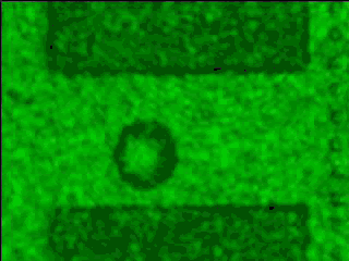
This series of four video clips (in dv format) shows how the presence of nanobubbles affects the growth of microbubbles on a microheater. Each frame of each 'movie' actually is from a different bubble nucleation and growth process. Each frame is taken after a set delay from the start of the voltage pulse to the heater. The frames are assembled into a movie in the order of increasing delay times. The flash duration of the laser is 7.5 nanoseconds. The distance across the microheater (top to bottom) is about 15 micrometers.First Nucleation: This clip shows bubble nucleation during the first pulse of a two-pulse sequence. The first frame is at a delay of 3.5 microseconds from the start of the voltage pulse. Succeeding frames are 10 ns apart. This clip in dv format. or This clip in avi format.
Note that although each frame is from a different bubble nucleation event, the images appear to be the steady growth of a single bubble, demonstrating the reproducibility of bubble growth during the first pulse.
This series of four video clips (in dv format) shows how the presence of nanobubbles affects the growth of microbubbles on a microheater. Each frame of each 'movie' actually is from a different bubble nucleation and growth process. Each frame is taken after a set delay from the start of the voltage pulse to the heater. The frames are assembled into a movie in the order of increasing delay times. The flash duration of the laser is 7.5 nanoseconds. The distance across the microheater (top to bottom) is about 15 micrometers.Growth and Collapse: during the first pulse of a two-pulse sequence. The first frame is at a delay of 3.5 microseconds from the start of the voltage pulse. Succeeding frames are 50 ns apart. This clip in dv format. or This clip in avi format.
This series of four video clips (in dv format) shows how the presence of nanobubbles affects the growth of microbubbles on a microheater. Each frame of each 'movie' actually is from a different bubble nucleation and growth process. Each frame is taken after a set delay from the start of the voltage pulse to the heater. The frames are assembled into a movie in the order of increasing delay times. The flash duration of the laser is 7.5 nanoseconds. The distance across the microheater (top to bottom) is about 15 micrometersSecond Nucleation: This clip shows bubble nucleation during the second pulse of a two-pulse sequence. The first frame is at a delay of 1.8 microseconds from the start of the voltage pulse. This clip in dv format. or This clip in avi format.
Succeeding frames are 10 ns apart. When measured from the start of their respective voltage pulses, bubbles from the second pulse appear 1.7 microseconds earlier than for the case of the first pulse. Note that because each frame is from a different bubble nucleation event, the growth process seems chaotic when viewed as a movie. This is evidence for the nature of the occurrence of the nucleation sites—nanobubbles formed at scattered locations from the collapse of the bubble from the first pulse.
This series of four video clips (in dv format) shows how the presence of nanobubbles affects the growth of microbubbles on a microheater. Each frame of each 'movie' actually is from a different bubble nucleation and growth process. Each frame is taken after a set delay from the start of the voltage pulse to the heater. The frames are assembled into a movie in the order of increasing delay times. The flash duration of the laser is 7.5 nanoseconds. The distance across the microheater (top to bottom) is about 15 micrometers.Growth and Collapse: during the second pulse of a two-pulse sequence. The first frame is at a delay of 1.8 microseconds from the start of the voltage pulse. Succeeding frames are 50 ns apart. This clip in dv format. or This clip in avi format.
Contact: Michael Baum michael.baum@nist.gov 301-975-2763 National Institute of Standards and Technology (NIST)

Technorati Tags: or and or and or and or and or and or and or and or

No comments:

Post a Comment