Tiny ion pump sets new standard in cooling hot computer chips
FULL TEXT, ion pump cooling hot computer chips.
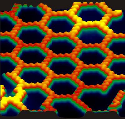
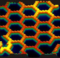 | Caption: Anthraquinone molecules form chains of molecules that weave themselves into a sheet of hexagons on a polished copper surface. Credit: Ludwig Bartels's research group, UCR, Usage Restrictions: None. |
FULL TEXT,
Honeycomb Network Comprised of Anthraquinone Molecules, Molecules spontaneously form honeycomb network featuring pores of unprecedented size
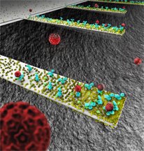
 | This rendition depicts an array of tiny, diving-boardlike devices called nanocantilevers. The devices are coated with antibodies to capture viruses, which are represented as red spheres. |
New findings about the behavior of the cantilevers could be crucial in designing a new class of ultra-small sensors for detecting viruses, bacteria and other pathogens. (Image generated by Seyet, LLC)
Nanocantilevers
High Resolution Image. FULL TEXT
'Nanocantilevers' yield surprises critical for designing new detectors.
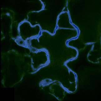
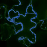 | This false-color image shows a cell from the epidermis of an Arabidopsis thaliana plant; the cell has been marked with fluorescent imaging sensors designed to detect the sugar glucose. |
In this image, only the densely packed interior of the cell in which most metabolic functions occur—called the cytosol—is targeted by the glucose sensors. The dark area sits inside the vacuole—a large storage organelle that can occupy up to 90% of the cell’s volume.
(Image courtesy Sylvie Lalonde and Wolf Frommer; click for higher resolution.) FULL TEXT
Sugar metabolism tracked in living plant tissues, in real time.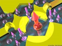 | Nanoscale metallic electrodes (in yellow) can be used to confine electrons in small regions, forming quantum dots. Two quantum dots connected to each other form a double quantum dot. |
In this case, one of the dots is in the Kondo state, in which the magnetic moment of the confined electron (large red arrow) is compensated (“screened”) by the magnetic moment of surrounding electrons, resulting in a zero net magnetic moment for the entire system. art by: Luis Dias/Ohio University. FULL TEXT,
Double Quantum Dots Control Kondo Effect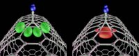 | Based on a new theory, MIT scientists may be able to manipulate carbon nanotubes -- |
one of the strongest known materials and one of the trickiest to work with -- without destroying their extraordinary electrical properties. FULL TEXT,
scientists tame tricky carbon nanotubes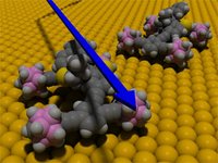 | This animation depicts two motorized nanocars on a gold surface. The nanocar consists of a rigid chassis and four alkyne axles that spin freely |
and swivel independently of one another. The wheels are spherical molecules of carbon, hydrogen and boron called p-carborane. FULL TEXT,
Nanocar inventor named top nanotech innovator | Caption: In NIST's Einstein-de Haas experiment. |
the movements of a cantilever were measured with an optical-fiber laser interferometer, The optical fiber is 125 micrometers in diameter, and the end is positioned less than 10 micrometers from the cantilever surface. Credit: Credit: John Moreland/NIST, Usage Restrictions: None. FULL TEXT,
Einstein's magnetic effect is measured on microscale


No comments:
Post a Comment