Saturday, January 26, 2013

Nanotubes the world's darkest known substance, to make an ultraefficient, highly accurate optical power detector

Nanotubes the world's darkest known substance, to make an ultraefficient, highly accurate optical power detector.

The National Institute of Standards and Technology (NIST) has demonstrated a novel chip-scale instrument made of carbon nanotubes that may simplify absolute measurements of laser power, especially the light signals transmitted by optical fibers in telecommunications networks.

The prototype device, a miniature version of an instrument called a cryogenic radiometer, is a silicon chip topped with circular mats of carbon nanotubes standing on end.* The mini-radiometer builds on NIST's previous work using nanotubes, the world's darkest known substance, to make an ultraefficient, highly accurate optical power detector,** and advances NIST's ability to measure laser power delivered through fiber for calibration customers.***


Caption: The circular patch of carbon nanotubes on a pink silicon backing is one component of NIST’s new cryogenic radiometer, shown with a quarter for scale. Gold coating and metal wiring has yet to be added to the chip. The radiometer will simplify and lower the cost of disseminating measurements of laser power.

Credit: Tomlin/NIST. Usage Restrictions: None.
"This is our play for leadership in laser power measurements," project leader John Lehman says. "This is arguably the coolest thing we've done with carbon nanotubes. They're not just black, but they also have the temperature properties needed to make components like electrical heaters truly multifunctional."

NIST and other national metrology institutes around the world measure laser power by tracing it to fundamental electrical units. Radiometers absorb energy from light and convert it to heat. Then the electrical power needed to cause the same temperature increase is measured. NIST researchers found that the mini-radiometer accurately measures both laser power (brought to it by an optical fiber) and the equivalent electrical power within the limitations of the imperfect experimental setup. The tests were performed at a temperature of 3.9 K, using light at the telecom wavelength of 1550 nanometers.

The tiny circular forests of tall, thin nanotubes called VANTAs ("vertically aligned nanotube arrays") have several desirable properties. Most importantly, they uniformly absorb light over a broad range of wavelengths and their electrical resistance depends on temperature.

The versatile nanotubes perform three different functions in the radiometer. One VANTA mat serves as both a light absorber and an electrical heater, and a second VANTA mat serves as a thermistor (a component whose electrical resistance varies with temperature). The VANTA mats are grown on the micro-machined silicon chip, an instrument design that is easy to modify and duplicate. In this application, the individual nanotubes are about 10 nanometers in diameter and 150 micrometers long.

By contrast, ordinary cryogenic radiometers use more types of materials and are more difficult to make. They are typically hand assembled using a cavity painted with carbon as the light absorber, an electrical wire as the heater, and a semiconductor as the thermistor. Furthermore, these instruments need to be modeled and characterized extensively to adjust their sensitivity, whereas the equivalent capability in NIST's mini-radiometer is easily patterned in the silicon.

NIST plans to apply for a patent on the chip-scale radiometer. Simple changes such as improved temperature stability are expected to greatly improve device performance. Future research may also address extending the laser power range into the far infrared, and integration of the radiometer into a potential multipurpose "NIST on a chip" device.
###

* N.A. Tomlin, J.H. Lehman. Carbon nanotube electrical-substitution cryogenic radiometer: initial results. Optics Letters. Vol. 38, No. 2. Jan. 15, 2013.

* N.A. Tomlin, J.H. Lehman. Carbon nanotube electrical-substitution cryogenic radiometer: initial results. Optics Letters. Vol. 38, No. 2. Jan. 15, 2013.

** See 2010 NIST Tech Beat article, "Extreme Darkness: Carbon Nanotube Forest Covers NIST's Ultra-dark Detector," at www.nist.gov/pml/div686/dark_081710.cfm.

***See 2011 NIST Tech Beat article, "Prototype NIST Device Measures Absolute Optical Power in Fiber at Nanowatt Levels," at www.nist.gov/pml/div686/radiometer-122011.cfm.

Contact: Laura Ost laura.ost@nist.gov 303-497-4880 National Institute of Standards and Technology (NIST)

Effective Passivation of Black Silicon Surfaces by Atomic Layer Deposition

Black silicon can take efficiency of solar cells to new levels

Scientists at Aalto University have demonstrated results that show a huge improvement in the light absorption and the surface passivation of silicon nanostructures. This has been achieved by applying atomic layer coating. The results advance the development of devices that require high sensitivity light response such as high efficiency solar cells.

- This method provides extremely good surface passivation. Simultaneously, it reduces the reflectance further at all wavelengths.These results are very promising considering the use of black silicon (b-Si) surfaces on solar cells to increase the efficiency to completely new levels, tells researcher scientist. Päivikki Repo.

More effective surface passivation methods than those used in the past have been needed to make black silicon a viable material for commercial applications. Good surface passivation is crucial in photonic applications such as solar cells. So far, the poor charge carrier transport properties attributed to nanostructured surfaces have been more detrimental for the final device operation than the gain obtained from the reduced reflectance.

Black Silicon

Black silicon (b-Si) can also be used in other technologies than solar cells. Numerous applications suggested for b-Si include drug analysis.

Black silicon has been a subject of great interest in various fields including photovoltaics for its ability to reduce the surface reflectance even below 1 per cent. However, many b-Si applications - especially solar cells - suffer from increased surface recombination resulting in poor spectral response. This is particularly problematic at short wavelengths.

The research has just been published in the Journal of Photovoltaics. The research is carried out by Aalto University, Finland, together with experts from Fraunhofer Institute for Solar Energy Systems ISE, Germany.

Further information:
Research Scientist Päivikki Repo, Aalto University School of Electrical Engineering
salla.repo@aalto.fi
tel. +358 504361156

Full citation: Effective Passivation of Black Silicon Surfaces by Atomic Layer Deposition, P. Repo, A. Haarahiltunen, L. Sainiemi, M. Yli-Koski, H. Talvitie, M. C. Schubert, and H. Savin pp. 90-94 in IEEE Journal of Photovoltaics, JPV January 2013. DOI: 10.1109/JPHOTOV.2012.2210031

Wednesday, January 23, 2013

Anomalous High Ionic Conductivity of Nanoporous ß-Li3PS4

OAK RIDGE, Tenn., Jan. 23, 2013 — Looking toward improved batteries for charging electric cars and storing energy from renewable but intermittent solar and wind, scientists at Oak Ridge National Laboratory have developed the first high-performance, nanostructured solid electrolyte for more energy-dense lithium ion batteries.

Today's lithium-ion batteries rely on a liquid electrolyte, the material that conducts ions between the negatively charged anode and positive cathode. But liquid electrolytes often entail safety issues because of their flammability, especially as researchers try to pack more energy in a smaller battery volume. Building batteries with a solid electrolyte, as ORNL researchers have demonstrated, could overcome these safety concerns and size constraints.

"To make a safer, lightweight battery, we need the design at the beginning to have safety in mind," said ORNL's Chengdu Liang, who led the newly published study in the Journal of the American Chemical Society. "We started with a conventional material that is highly stable in a battery system - in particular one that is compatible with a lithium metal anode."

lithium thiophosphate

ORNL researchers developed a nanoporous solid electrolyte (bottom left and in detail on right) from a solvated precursor (top left). The material conducts ions 1,000 times faster than its natural bulk form and enables more energy-dense lithium ion batteries.
The ability to use pure lithium metal as an anode could ultimately yield batteries five to 10 times more powerful than current versions, which employ carbon based anodes.

"Cycling highly reactive lithium metal in flammable organic electrolytes causes serious safety concerns," Liang said. "A solid electrolyte enables the lithium metal to cycle well, with highly enhanced safety."

The ORNL team developed its solid electrolyte by manipulating a material called lithium thiophosphate so that it could conduct ions 1,000 times faster than its natural bulk form. The researchers used a chemical process called nanostructuring, which alters the structure of the crystals that make up the material.

"Think about it in terms of a big crystal of quartz vs. very fine beach sand," said coauthor Adam Rondinone. "You can have the same total volume of material, but it's broken up into very small particles that are packed together.
It's made of the same atoms in roughly the same proportions, but at the nanoscale the structure is different. And now this solid material conducts lithium ions at a much greater rate than the original large crystal."

The researchers are continuing to test lab scale battery cells, and a patent on the team's invention is pending.

"We use a room-temperature, solution-based reaction that we believe can be easily scaled up," Rondinone said. "It's an energy-efficient way to make large amounts of this material."

For information about industry collaboration opportunities, please visit the ORNL Partnerships website at http://www.ornl.gov/adm/partnerships/index.shtml.

The study is published as "Anomalous High Ionic Conductivity of Nanoporous ß-Li3PS4," and its ORNL coauthors are Zengcai Liu, Wujun Fu, Andrew Payzant, Xiang Yu, Zili Wu, Nancy Dudney, Jim Kiggans, Kunlun Hong, Adam Rondinone and Chengdu Liang. The work was sponsored by the Division of Materials Sciences and Engineering in DOE's Office of Science.

The materials synthesis and characterization were supported by the Center for Nanophase Materials Sciences at ORNL. CNMS is one of the five DOE Nanoscale Science Research Centers supported by the DOE Office of Science, premier national user facilities for interdisciplinary research at the nanoscale. Together the NSRCs comprise a suite of complementary facilities that provide researchers with state-of-the-art capabilities to fabricate, process, characterize and model nanoscale materials, and constitute the largest infrastructure investment of the National Nanotechnology Initiative. The NSRCs are located at DOE's Argonne, Brookhaven, Lawrence Berkeley, Oak Ridge and Sandia and Los Alamos national laboratories.

DOE's Office of Science is the single largest supporter of basic research in the physical sciences in the United States, and is working to address some of the most pressing challenges of our time. For more information, please visit science.energy.gov.

Contact: Morgan McCorkle mccorkleml@ornl.gov 865-574-7308 DOE/Oak Ridge National Laboratory

Thursday, January 17, 2013

Telechelic Star Polymers as Self-Assembling Units from the Molecular to the Macroscopic Scale

Telechelic Star Polymers as Self-Assembling Units from the Molecular to the Macroscopic Scale.

Barbara Capone of the Computational Physics Group of the University of Vienna has developed a new method for the construction of building blocks at the nanoscale. The researcher in Soft Matter Physics, who works at the group of Christos Likos, Professor for Multiscale Computational Physics, is specialized on topics of self-assembly of materials at the nanoscale and she has published, together with her colleagues, a paper at the prestigious Journal "Physical Review Letters" on "soft Lego".

In developing these novel self-assembling materials, postdoc Barbara Capone has focused on the design of organic and inorganic building blocks, which are robust and can be produced at large scale. Capone has put forward, together with her colleagues at the Universities of Vienna and Mainz, a completely new pathway for the construction of building blocks at the nanoscale.

"Soft Lego" orders in crystal structures

Telechelic Star Polymers

Simulation snapshot of a diamond crystal built of soft patchy diblock star polymers (Copyright: American Physical Society)
The team of researchers has shown that so-called block copolymer stars – that means polymers that consist of two different blocks and they are chemically anchored on a common point – have a robust and flexible architecture and they possess the ability to self-assemble at different levels. At the single-molecule level, they first order as soft patchy colloids which serve then as "soft Lego" for the emergence of larger structures. At the next level of self-assembly, the colloids form complex crystal structures, such as diamond or cubic phases.

The spatial ordering in the crystals can be steered through the architecture of the "soft Lego" and opens up the possibility for the construction of new materials at the macroscopic scale with desired structure. In this way, crystals can be built that have applications in, e.g., photonics, acting as filters for light of certain frequencies or as light guides.

Contact: Barbara Capone barbara.capone@univie.ac.at 43-142-777-3236 University of Vienna

Tuesday, January 15, 2013

Asbestos-like Pathogenicity of Long Carbon Nanotubes Alleviated by Chemical Functionalization

Asbestos-like Pathogenicity of Long Carbon Nanotubes Alleviated by Chemical Functionalization.

Safety fears about carbon nanotubes, due to their structural similarity to asbestos, have been alleviated following research showing that reducing their length removes their toxic properties.

In a new study, published today in the journal Angewandte Chemie, evidence is provided that the asbestos-like reactivity and pathogenicity reported for long, pristine nanotubes can be completely alleviated if their surface is modified and their effective length is reduced as a result of chemical treatment.

First atomically described in the 1990s, carbon nanotubes are sheets of carbon atoms rolled up into hollow tubes just a few nanometres in diameter. Engineered carbon nanotubes can be chemically modified, with the addition of chemotherapeutic drugs, fluorescent tags or nucleic acids – opening up applications in cancer and gene therapy.

Furthermore, these chemically modified carbon nanotubes can pierce the cell membrane, acting as a kind of 'nano-needle', allowing the possibility of efficient transport of therapeutic and diagnostic agents directly into the cytoplasm of cells.

Among their downsides however, have been concerns about their safety profile. One of the most serious concerns, highlighted in 2008, involves the carcinogenic risk from the exposure and persistence of such fibres in the body. Some studies indicate that when long untreated carbon nanotubes are injected to the abdominal cavity of mice they can induce unwanted responses resembling those associated with exposure to certain asbestos fibres.

ext

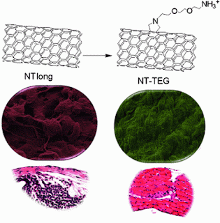
Sometimes shorter is better: The apparent similarity between multi-walled carbon nanotubes (MWNTs) and asbestos fibers has generated serious concerns about their safety profile. The asbestos-like pathogenicity observed for long, pristine nanotubes (NTlong, see scheme) can be completely alleviated if their effective length is decreased as a result of chemical functionalization, such as with tri(ethylene glycol) (TEG).
In this paper, the authors describe two different reactions which ask if any chemical modification can render the nanotubes non-toxic. They conclude that not all chemical treatments alleviate the toxicity risks associated with the material. Only those reactions that are able to render carbon nanotubes short and stably suspended in biological fluids without aggregation are able to result in safe, risk-free material.

Professor Kostas Kostarelos, Chair of Nanomedicine at the UCL School of Pharmacy who led the research with his long term collaborators Doctor Alberto Bianco of the CNRS in Strasbourg, France and Professor Maurizio Prato of the University of Trieste, Italy, said: "The apparent structural similarity between carbon nanotubes and asbestos fibres has generated serious concerns about their safety profile and has resulted in many unreasonable proposals of a halt in the use of these materials even in well-controlled and strictly regulated applications, such as biomedical ones. What we show for the first time is that in order to design risk-free carbon nanotubes both chemical treatment and shortening are needed."

He added: "Creative strategies to identify the characteristics that nanoparticles should possess in order to be rendered 'safe-for-use', and the ways to achieve that, are essential as nanotechnology and its tools are maturing into applications and becoming part of our everyday lives."

###

About UCL (University College London)

Founded in 1826, UCL was the first English university established after Oxford and Cambridge, the first to admit students regardless of race, class, religion or gender and the first to provide systematic teaching of law, architecture and medicine.

We are among the world's top universities, as reflected by our performance in a range of international rankings and tables. According to the Thomson Scientific Citation Index, UCL is the second most highly cited European university and the 15th most highly cited in the world.

UCL has nearly 25,000 students from 150 countries and more than 9,000 employees, of whom one third are from outside the UK. The university is based in Bloomsbury in the heart of London, but also has two international campuses – UCL Australia and UCL Qatar. Our annual income is more than £800 million.

Contact: Clare Ryan clare.ryan@ucl.ac.uk 44-020-310-83846 University College London

For more information, please contact Professor Kostas Kostarelos on tel: +44 (0)207 753 5956, email: k.kostarelos@ucl.ac.uk

Thursday, January 10, 2013

Molecular Peptide Synthesizer could hold key to more efficient manufacturing

An industrial revolution on a minute scale is taking place in laboratories at The University of Manchester with the development of a highly complex machine that mimics how molecules are made in nature.

The artificial molecular machine developed by Professor David Leigh FRS and his team in the School of Chemistry is the most advanced molecular machine of its type in the world. Its development has been published in the journal Science.

Professor Leigh explains: "The development of this machine which uses molecules to make molecules in a synthetic process is similar to the robotic assembly line in car plants. Such machines could ultimately lead to the process of making molecules becoming much more efficient and cost effective. This will benefit all sorts of manufacturing areas as many manmade products begin at a molecular level. For example, we're currently modifying our machine to make drugs such as penicillin."

The machine is just a few nanometres long (a few millionths of a millimetre) and can only be seen using special instruments. Its creation was inspired by natural complex molecular factories where information from DNA is used to programme the linking of molecular building blocks in the correct order. The most extraordinary of these factories is the ribosome, a massive molecular machine found in all living cells.


Professor Leigh’s molecular machine is based on the ribosome. It features a functionalized nanometre-sized ring that moves along a molecular track, picking up building blocks located on the path and connecting them together in a specific order to synthesize the desired new molecule. First the ring is threaded onto a molecular strand using copper ions to direct the assembly process. Then a “reactive arm” is attached to the rest of the machine and it starts to operate.

The ring moves up and down the strand until its path is blocked by a bulky group. The reactive arm then detaches the obstruction from the track and passes it to another site on the machine, regenerating the active site on the arm. The ring is then free to move further along the strand until its path is obstructed by the next building block. This, in turn, is removed and passed to the elongation site on the ring, thus building up a new molecular structure on the ring. Once all the building blocks are removed from the track, the ring de-threads and the synthesis is over.

Credit: Miriam Wilson. Usage Restrictions: Please credit Miriam Wilson.
Professor Leigh's machine is based on the ribosome. It features a functionalized nanometre-sized ring that moves along a molecular track, picking up building blocks located on the path and connecting them together in a specific order to synthesize the desired new molecule.

First the ring is threaded onto a molecular strand using copper ions to direct the assembly process. Then a "reactive arm" is attached to the rest of the machine and it starts to operate. The ring moves up and down the strand until its path is blocked by a bulky group. The reactive arm then detaches the obstruction from the track and passes it to another site on the machine, regenerating the active site on the arm. The ring is then free to move further along the strand until its path is obstructed by the next building block. This, in turn, is removed and passed to the elongation site on the ring, thus building up a new molecular structure on the ring. Once all the building blocks are removed from the track, the ring de-threads and the synthesis is over.

Professor Leigh says the current prototype is still far from being as efficient as the ribosome: "The ribosome can put together 20 building blocks a second until up to 150 are linked. So far we have only used our machine to link together 4 blocks and it takes 12 hours to connect each block. But you can massively parallel the assembly process: We are already using a million million million (1018) of these machines working in parallel in the laboratory to build molecules."

Professor Leigh continues: "The next step is to start using the machine to make sophisticated molecules with more building blocks. The potential is for it to be able to make molecules that have never been seen before. They're not made in nature and can't be made synthetically because of the processes currently used. This is a very exciting possibility for the future."

###

Contact: Morwenna Grills Morwenna.Grills@manchester.ac.uk
44-161-275-2111 University of Manchester

Wednesday, January 09, 2013

Investigation of the Piezoelectric Effect as a Means to Generate X-Rays

COLUMBIA, Mo. — The hand-held scanners, or tricorders, of the Star Trek movies and television series are one step closer to reality now that a University of Missouri engineering team has invented a compact source of X-rays and other forms of radiation. The radiation source, which is the size of a stick of gum, could be used to create inexpensive and portable X-ray scanners for use by doctors, as well as to fight terrorism and aid exploration on this planet and others.

“Currently, X-ray machines are huge and require tremendous amounts of electricity,” said Scott Kovaleski, associate professor of electrical and computer engineering at MU. “In approximately three years, we could have a prototype hand-held X-ray scanner using our invention. The cell-phone-sized device could improve medical services in remote and impoverished regions and reduce health care expenses everywhere.”

Kovaleski suggested other uses for the device. In dentists’ offices, the tiny X-ray generators could be used to take images from the inside of the mouth shooting the rays outward, reducing radiation exposure to the rest of the patients’ heads. At ports and border crossings, portable scanners could search cargoes for contraband, which would both reduce costs and improve security. Interplanetary probes, like the Curiosity rover, could be equipped with the compact sensors, which otherwise would require too much energy.

compact radiation source

The compact radiation source developed by Kovaleski's team CREDIT: Peter Norgard, University of Missouri.
The accelerator developed by Kovaleski’s team could be used to create other forms of radiation in addition to X-rays. For example, the invention could replace the radioactive materials, called radioisotopes, used in drilling for oil as well as other industrial and scientific operations. Kovaleski’s invention could replace radioisotopes with a safer source of radiation that could be turned off in case of emergency.

“Our device is perfectly harmless until energized, and even then it causes relatively low exposures to radiation,” said Kovaleski. “We have never really had the ability to design devices around a radioisotope with an on-off switch. The potential for innovation is very exciting.”

The device uses a crystal to produce more than 100,000 volts of electricity from only 10 volts of electrical input with low power consumption. Having such a low need for power could allow the crystal to be fueled by batteries. The crystal, made from a material called lithium niobate, uses the piezoelectric effect to amplify the input voltage. Piezoelectricity is the phenomenon whereby certain materials produce an electric charge when the material is under stress.

Kovaleski’s team published “Investigation of the Piezoelectric Effect as a Means to Generate X-Rays” in the journal IEEE Transaction on Plasma Science. Kovaleski is interim department chair of the Electrical & Computer Engineering in MU’s Department of Engineering. --30--

Jan. 08, 2013. Story Contact(s): Timothy Wall, walltj@missouri.edu, 573-882-3346


Sunday, January 06, 2013

Optofluidics allow for a new understanding of resistance to antibiotics

Rethinking bacterial persistence. Optofluidics allow for a new understanding of resistance to antibiotics

It's often difficult to completely eliminate a bacterial infection with antibiotics; part of the population usually manages to survive. We've known about this phenomenon for quite some time, dating back nearly to the discovery of penicillin. For more than 50 years, scientists have believed that the resistant bacteria were individuals that had stopped growing and dividing.

Up to now, in fact, it hasn't been possible to track the growth of cells before and after their exposure to antibiotics, which makes any analysis of the phenomenon quite imprecise. "Using microfluidics, we can now observe every bacterium individually, instead of having to count a population," says John McKinney, director of EPFL's Microbiology and Microsystems Laboratory (LMIC).

Active survivors


Bacterial ife, growth and death is now to be seen more accurately. © 2012 LMIC / EPFL
This new tool has revealed quite a few surprises. "We thought that surviving bacteria made up a fixed population that stopped dividing, but instead we found that some of them continued to divide and others died. The persistent population is thus very dynamic, and the cells that constitute it are constantly changing – even though the total number of cells remains the same. Because they're dividing, the bacteria can mutate and thus develop resistance in the presence of the antibiotic," explains LMIC scientist Neeraj Dhar.

This point is extremely important. "We were able to eliminate a purely genetic explanation of the phenomenon," continues Dhar. In other words, "a population of genetically identical bacteria consists of individuals with widely varying behavior. Some of them can adapt to stressors that they have not previously encountered, thanks to the selection of persistent individuals. This could lead to a revision of the entire theory of adaptation," says McKinney.

Intermittent efficiency

The EPFL scientists were particularly interested in a relative of the tuberculosis bacterium. Their observations enabled them to formally challenge the argument that persistent bacteria are those that have stopped growing and dividing. "We were able to reveal the role of an enzyme whose presence is necessary in order for the antibiotic to work, and show that the bacilli produced this enzyme in a pulsatile and random manner," explains Dhar. "Our measurements showed that bacterial death correlated more closely with the expression of this enzyme than with their growth factor." The research is being published this week in Science magazine.

These conclusions could mark the beginning of a new theory of bacterial resistance, or perhaps even introduce a new view of how such resistance evolves. Further research is being done using other microorganisms, such as tuberculosis and E. coli bacteria. The persistence of some cancer cells to treatment could also be studied in a different manner. "It's a new approach for trying to figure out why some infections are so difficult to eliminate. The techniques we've developed for this study are now also being used to develop new antibiotics, in collaboration with pharmaceutical companies," says McKinney, adding that "it is the microengineering expertise at EPFL that has enabled us to create these innovative tools and open up new avenues for investigation."

###

Contact: Emmanuel Barraud emmanuel.barraud@epfl.ch 41-796-283-642 Ecole Polytechnique Fédérale de Lausanne

Thursday, January 03, 2013

Transient Enhancement and Spectral Narrowing of The Photothermal Effect of Plasmonic Nanoparticles Under Pulsed Excitation

Transient Enhancement and Spectral Narrowing of The Photothermal Effect of Plasmonic Nanoparticles Under Pulsed Excitation - Rice University researchers show short laser pulses selectively heat gold nanoparticles.

HOUSTON – (Jan. 3, 2013) – Plasmonic gold nanoparticles make pinpoint heating on demand possible. Now Rice University researchers have found a way to selectively heat diverse nanoparticles that could advance their use in medicine and industry.

Rice scientists led by Dmitri Lapotko and Ekaterina Lukianova-Hleb showed common gold nanoparticles, known since the 19th century as gold colloids, heat up at near-infrared wavelengths as narrow as a few nanometers when hit by very short pulses of laser light. The surprising effect reported in Advanced Materials appears to be related to nonstationary optical excitation of plasmonic nanoparticles. Plasmons are free electrons on the surface of metals that become excited by the input of energy, typically from light. Moving plasmons can transform optical energy into heat.

"The key idea with gold nanoparticles and plasmonics in general is to convert energy," Lapotko said. "There are two aspects to this: One is how efficiently you can convert energy, and here gold nanoparticles are world champions. Their optical absorbance is about a million times higher than any other molecules in nature.


Different types of nanoparticles – in this case, shells, rods and solid spheres – mixed together can be activated individually with pulsed laser light at different wavelengths, according to researchers at Rice University. The tuned particles' plasmonic response, enhanced by nanobubbles that form at the surface, can be narrowed to a few nanometers under a spectroscope and are easily distinguishable from each other. (Credit: Lapotko Group/Rice University)


Rice University researchers found that pulsed (or "nonstationary ") lasers could narrow the response spectra of 60-nanometer-wide gold nanoshells to a very narrow spectral band (red peak), as opposed to continuous ("stationary") excitation by laser (green peak). The discovery opens new possibilities for the use of metallic nanoparticles in medical and electronic applications. Credit: Lapotko Group/Rice University. Usage Restrictions: None


The strong response of plasmonic gold nanoparticles to pulsed ("nonstationary") lasers rather than continuous ("stationary") excitation by lasers appears to be due to the influence of nanobubbles on the particles, according to researchers at Rice University. (Credit: Lapotko Group/Rice University)


"The second aspect is how precisely one can use laser radiation to make this photothermal conversion happen," he said. Particles traditionally respond to wide spectra of light, and not much of it is in the valuable near-infrared region. Near-infrared light is invisible to water and, more critically for biological applications, to tissue.

"This was the problem," Lapotko said. "All nanoparticles, beginning with solid gold colloids and moving to more sophisticated, engineered gold nanoshells, nanorods, cages and stars, have very wide spectra, typically about 100 nanometers, which means we were allowed to use only one type of nanoparticle at a time. If we tried to use different types, their spectra overlapped and we did not benefit from the high tunability of lasers."

The discovery allows controlled laser pulses to tune the absorbance spectrum of plain gold colloids, Lapotko said. "This novel approach is counter to the established paradigm that assumes optical properties of nanoparticles are pre-set during their fabrication and stay constant during their optical excitation," he said.

The Rice lab showed basic colloidal gold nanoparticles could be efficiently activated by a short laser pulse at 780 nanometers, with an 88-fold amplification of the photothermal effect seen with a continuous laser. The researchers repeated their experiment with nanoparticle clusters in water, in living cancer cells and in animals, with the same or better results: they showed spectral peaks two nanometers wide. Such narrow photothermal spectra had never been seen for metal nanoparticles, either singularly or in clusters.

The effect appears to depend on vapor nanobubbles that form when the particles heat liquid in their immediate environment. The nanobubbles grow and burst in an instant. "Instead of using the nanoparticle as a heat sink with a continuous, stationary laser, we're creating a transient, nonstationary situation in which the particle interacts with the incident laser in a totally different way," Lapotko said. He said the effect is repeatable and works with laser pulses shorter than 100 picoseconds.

Even better, an experiment with mixed nanorods and nanoshells found they responded to laser pulses with strong, distinct signals at wavelengths 10 nanometers apart. That means two or more types of nanoparticles in the same location can be selectively activated on demand.

"The nanoparticles we used were nothing fancy; they were used in the 19th century by Michael Faraday, and it was believed they could do nothing in the near-infrared," he said. "That was the major motivation for people to invent nanorods, nanoshells and the other shapes. Here, we prove these inexpensive particles can behave quite well in near-infrared." He said the discovery opens the possibility that many metal nanoparticles could be used in biomedical and industrial applications where spectral selectivity and tuning would provide "unprecedented" precision.

"This is still more a phenomenon rather than a firmly established mechanism, with a nice theoretical basis," Lapotko said. "But when fully clarified, it could become a universal tool."

###

Co-authors of the paper are Alexey Volkov, a research scientist at the University of Virginia, and Xiangwei Wu, an associate professor in the Department of Head and Neck Surgery at the University of Texas MD Anderson Cancer Center. Lapotko is a faculty fellow in biochemistry and cell biology, and Lukianova-Hleb is a research scientist at Rice.

The National Institutes of Health supported the research.

Contact: Mike Williams mikewilliams@rice.edu 713-348-6728 Rice University

Tuesday, January 01, 2013

Geometric and topological analyses of micro-materials in three-dimensional liquid crystal experiments

Liquid crystal research, future applications advance - Mathematics explains the observed 'beautiful and complex patterns revealed' in three-dimensional liquid crystal experiments, expected to lead to creation of new materials that can be actively controlled.

AMHERST, Mass. – Contributing geometric and topological analyses of micro-materials, University of Massachusetts Amherst mathematician Robert Kusner aided experimental physicists at the University of Colorado (UC) by successfully explaining the observed "beautiful and complex patterns revealed" in three-dimensional liquid crystal experiments. The work is expected to lead to creation of new materials that can be actively controlled.

Kusner is a geometer, an expert in the analysis of variational problems in low-dimensional geometry and topology, which concerns properties preserved under continuous deformation such as stretching and bending. His work over 3 decades has focused on the geometry and topology of curves, surfaces and other spaces that arise in nature, such as soap films, knots and the shapes of fluid droplets. Kusner agrees with physicist and lead author Ivan Smalyukh of UC Boulder that their collaboration is the first to show in experiments that some of the most fundamental topological theorems hold up in real materials. Their findings appear in the current early online issue of Nature.

Polarized Light in a Liquid Crystal
This image shows polarized light interacting with a particle injected into a liquid crystal medium. Credit: Bohdan Senyuk and Ivan Smalyukh, Colorado University. Usage Restrictions: None. Related news release: Liquid crystal research, future applications advance
UMass Amherst's Kusner explains, "There are two important aspects of this work. First, the experimental work by the Colorado team, who fabricated topologically complex micro-materials allowing controlled experiments of three-dimensional liquid crystals. Second, the theoretical work performed by us mathematicians and theoretical physicists while visiting the University of California Santa Barbara's Kavli Institute for Theoretical Physics (KITP). We provided the geometric and topological analysis of these experiments, to explain the observed patterns and predict what patterns should be seen when experimental conditions are changed."

Kusner was the lone mathematician among four organizers of last summer's workshop on "Knotted Fields" at KITP, which led to this work. The workshop engaged about a dozen other mathematicians and about twice as many theoretical and experimental physicists in a month-long investigation of the interplay between low-dimensional topology and what physicists call "soft matter."

In their experiments, the physicists at UC Boulder showed that tiny topological particles injected into a liquid crystal medium behave in a manner consistent with established theorems in geometry and topology, Kusner says. The researchers say they have thus identified approaches for building new materials using topology.

UC Boulder's Smalyukh and colleagues set up the experiment by first creating colloids, solutions in which tiny particles are dispersed but not dissolved in a host medium, such as milk, paint and shaving cream. Specifically, they injected tiny, different-shaped particles into a liquid crystal, which behaves something like a liquid and a solid. Once injected into a liquid crystal, the particles behaved as predicted by topology.

Smalyukh says, "Our study shows that interaction between particles and molecular alignment in liquid crystals follows the predictions of topological theorems, making it possible to use these theorems in designing new composite materials with unique properties that cannot be encountered in nature or synthesized by chemists. These findings lay the groundwork for new applications in experimental studies of low-dimensional topology, with important potential ramifications for many branches of science and technology."

For example, he adds, these topological liquid crystal colloids could be used to upgrade current liquid crystal displays like those used in laptops and television screens, to allow them to interact with light in new, more energy efficient ways.

###

Besides Kusner at UMass Amherst and Smalyukh's group at UC Boulder, other investigators for this study are Sailing He of Zhejiang University, China and Randall Kamien and Tom Lubensky at the University of Pennsylvania.

Contact: Robert Kusner kusner@math.umass.edu 413-545-6022 University of Massachusetts at Amherst