Monday, October 31, 2011

World’s most advanced extreme-ultraviolet (EUV) microscope, SHARP (Semiconductor High-NA Actinic Reticle Review Project)

Moore’s Law, hardly a law but undeniably a persistent trend, says that every year and a half, the number of transistors that fit on a chip roughly doubles. It’s why electronics – from smart phones to flat screens, from MP4 players to movie cameras, from tablets to supercomputers – grow ever more varied, powerful, and compact, but also ever less expensive. Whether the trend can continue until it runs up against immutable laws of nature, like the finite size of an atom, depends on how far scientists and technicians can push electronic technologies down into the nanoworld with better tools for using short-wavelength light.

Now scientists at the U.S. Department of Energy’s Lawrence Berkeley National Laboratory (Berkeley Lab) have partnered with colleagues at leading semiconductor manufacturers to create the world’s most advanced extreme-ultraviolet (EUV) microscope. Called SHARP (a succinct acronym for a long name, the Semiconductor High-NA Actinic Reticle Review Project), the new microscope will be dedicated to photolithography, the central process in the creation of computer chips.

The $4.1 million, 1.5-year project will be led by Kenneth Goldberg of the Center for X-Ray Optics (CXRO) in Berkeley Lab’s Materials Science Division (MSD). Initially SHARP will be used in parallel with operations at the existing microscope on beamline 11.3.2 of Berkeley Lab’s Advanced Light Source (ALS). By the last quarter of 2012 the new EUV photomask-imaging microscope will replace the beamline’s aging facilities.

“EUV light is tricky to work with,” says Goldberg, “because every material absorbs it so strongly. So instead of glass lenses, EUV optical systems rely mainly on specialized mirrors with atomic-scale smoothness, topped by multilayer coatings for high reflectivity.” To maintain efficiency, the entire optical system has to be placed in a high-vacuum environment.

extreme ultraviolet (EUV) absorbing layer

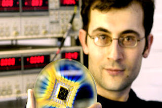
Kenneth Goldberg is seen in the reflective coating of a photolithography mask, contained in the clear plastic box, which he’s about to measure at the Advanced Light Source’s beamline 11.3.2. Inset at lower right shows a mask’s extreme-ultraviolet (EUV) absorbing layer, printed on a six-inch square of glass coated with multiple layers of molybdenum and silicon only billionths of a meter thick to reflect unwanted EUV. The patterned layer represents one level of a working microprocessor or memory chip, which may have 20 or more such levels. Its structures are less than one ten-millionth of a meter across and diffract visible light in rainbow patterns.
While the existing eight-year-old microscope at beamline 11.3.2, dubbed the AIT (for Actinic Inspection Tool), has unique imaging capabilities, the fast-moving nature of semiconductor technology means its future is limited. SHARP will exceed its performance in every metric: resolution, speed, uniformity of illumination, and coherence control. SHARP will enable forward-looking research years before commercial tools become available.

Within a few years, semiconductor devices will be measured in dimensions of 16, 11, or 8 nanometers, mere billionths of a meter. To mass-produce them, industry is pushing a photolithography process that uses EUV light with a wavelength of just 13.5 nanometers, 40 times smaller than visible light.

“At this short wavelength, we can print and image circuit patterns at nanometer length scales,” says Goldberg. “The new microscope will leverage years of cutting-edge EUV and soft-x‑ray microscopy experience, experimental systems-engineering at CXRO, and EUV optics expertise developed as part of the lithography research programs here.”

Goldberg says that the ALS, as one of the world’s brightest sources of EUV light, “is a great place to develop EUV lithography technologies.”

In lithography, photomasks are the key to mass production. A series of photomasks carry the master circuit patterns that are transferred onto a chip, layer by layer, to create working semiconductor devices. The masks are analogous to the negatives in a photographer’s darkroom, or master pages on a photocopier.

Minute imperfections or tiny particles of dust on a master, if not found and cleaned or fixed, ultimately cause chips to fail. Goldberg and his team have shown that defects and patterns can appear very different when viewed with non-EUV inspection tools such as electron microscopes, making EUV microscopy essential for the development of EUV masks because only in this way can damaging dust particles and other defects be identified reliably.

“Other microscopes can have wonderfully high resolution, but they can’t detect the wavelength-specific EUV response of mask patterns and defects,” says Goldberg, “and that’s necessary to make successful repairs.”

SHARP is called an “actinic” microscope because it uses the same EUV wavelengths used in production. Thus the new EUV microscope will enable semiconductor company researchers to better evaluate defects and repair strategies, mask materials and architectures, and advanced pattern features.

Like its predecessor, the SHARP microscope will also feature an array of lenses, side by side, so users can select the different imaging properties they need, much as a common lab microscope mounts different lenses on a rotating turret.

The high-magnification objective lenses for the new microscope are holographic Fresnel zoneplate lenses, microscopic objects produced by CXRO’s Nanowriter. The Nanowriter, under the direction of Erik Anderson, holds the world record for creating the highest resolution zoneplates for many synchrotron and other short-wavelength applications. The lenses are only slightly wider than a single human hair, yet they project high-quality images of the mask surface with up to 2,000 times magnification.

A special feature of the new microscope will be illumination coherence control. The ALS produces an EUV beam with laser-like coherence, ideal for many experiments. For microscopy, however, the image resolution can be improved by a factor of two by carefully re-engineering the illumination into a state called partial coherence. Microscopists have recognized the importance of partial coherence for years, and the synchrotron community is now catching up.

An angle-scanning mirror in the new microscope’s beamline illuminator will take the highly-coherent ALS light and steer it into patterns, like a mini-laser-light show, breaking and re-shaping the coherence properties. In this way, the SHARP microscope will replicate the properties of current and future tools for lithography production and research, giving researchers the most advanced look at what’s to come.

Additional information

With sales of some $51 billion a year, semiconductors are the United States’ second largest export product. Developing technology to produce and test the next generation computer chips is one of the industry’s core missions. For over a decade, semiconductor companies have sponsored photolithography-related research at Berkeley Lab through the Center for X-Ray Optics, including world-leading programs in optics, masks, and materials — most conducted on three CXRO beamlines at the Advanced Light Source.

Contact: Paul Preuss paul_preuss@lbl.gov 510-486-6249 DOE / Lawrence Berkeley National Laboratory

Lawrence Berkeley National Laboratory addresses the world’s most urgent scientific challenges by advancing sustainable energy, protecting human health, creating new materials, and revealing the origin and fate of the universe. Founded in 1931, Berkeley Lab’s scientific expertise has been recognized with 13 Nobel prizes. The University of California manages Berkeley Lab for the U.S. Department of Energy’s Office of Science. For more, visit www.lbl.gov.

Sunday, October 30, 2011

Nanotechnology and the Future of Electronics VIDEO

Dr. Robert J Trew
Alton and Mildred Lancaster Distinguished Professor
| Primary Research Interests

Nanoelectronics and Photonics (Including III - V Materials and Devices, Nanotechnology, Optical Materials and Photonic Devices, Quantum Engineering, Silicon Devices and Fabrication)

| Other Research Interests

Electronic Circuits and Systems (Including Electromagnetic Fields / Antenna Analysis, Microwave Devices and Circuits)
Power Electronics and Power Systems (Including Power Semiconductor Devices)

| Awards and Honors

2011 - American Association for the Advancement of Science (AAAS) Fellow
2006 - Distinguished Service Award for Service as a 2003-2005 IEEE Microwave Distinguished Lecturer
2006 - Named Editor-in-Chief of IEEE Proceedings for 2007-2009
2005 - Distinguished Service Award for Service as 2004 President of the IEEE Microwave Theory and Techniques Society ADCOM
2004 - Meritorious Service Award for Service as 2001-2003 Editor-in-Chief of IEEE Microwave Magazine
2004 - Named Distinguished Fellow, National Institute of Aerospace, NASA - Langley Research Center
2003 - Engineering Alumni Society Merit Award in Electrical Engineering, College of Engineering, University of Michigan
2003 - Named Alton and Mildred Lancaster Distinguished Professor of Electrical and Computer Engineering, NC State


2001 - Distinguished Service Award for Service as a 1997-2000 IEEE Microwave Distinguished Lecturer
2001 - Harry Diamond Memorial Award, Institute of Electrical and Electronics Engineers (IEEE-USA)
2001 - Named Willis G Worcester Distinguished Professor of Electrical and Computer Engineering, Virginia Tech
2000 - Hero of the US Army Research Office
2000 - IEEE Third Millennium Medal Award
1999 - Distinguished Service Award for Service as 1995-1997 Editor-in-Chief for IEEE Transactions on Microwave Theory and Techniques
1998 - Distinguished Educator Award, IEEE Microwave Theory & Techniques Society
1993 - Named George S Dively Distinguished Professor of Engineering, Case Western Reserve University
1993 - University Distinguished Speaker, Texas A&M University, College Station
1992 - ALCOA Foundation Distinguished Engineering Research Award (NC State)
1991 - Distinguished Scholarly Achievement Award (NC State)
1991 - IEEE Fellow

VIDEO CREDIT: ecencsu

TEXT CREDIT: The Department of Electrical and Computer Engineering at NC State University

Saturday, October 29, 2011

Merging of plasmonics and nanophotonics is promising the emergence of quantum information systems

WEST LAFAYETTE, Ind. – The merging of two technologies under development - plasmonics and nanophotonics - is promising the emergence of new "quantum information systems" far more powerful than today's computers.

The technology hinges on using single photons – the tiny particles that make up light – for switching and routing in future computers that might harness the exotic principles of quantum mechanics.

The quantum information processing technology would use structures called "metamaterials," artificial nanostructured media with exotic properties.

The metamaterials, when combined with tiny "optical emitters," could make possible a new hybrid technology that uses "quantum light" in future computers, said Vladimir Shalaev, scientific director of nanophotonics at Purdue University's Birck Nanotechnology Center and a distinguished professor of electrical and computer engineering.

The concept is described in an article to be published Friday (Oct. 28) in the journal Science. The article will appear in the magazine's Perspectives section and was written by Shalaev and Zubin Jacob, an assistant professor of electrical and computer engineering at the University of Alberta, Canada.

"A seamless interface between plasmonics and nanophotonics could guarantee the use of light to overcome limitations in the operational speed of conventional integrated circuits," Shalaev said.

quantum information systems

Structures called "metamaterials" and the merging of two technologies under development are promising the emergence of new "quantum information systems" far more powerful than today's computers. The concept hinges on using single photons – the tiny particles that make up light – for switching and routing in future computers that might harness the exotic principles of quantum mechanics. The image at left depicts a "spherical dispersion" of light in a conventional material, and the image at right shows the design of a metamaterial that has a "hyperbolic dispersion" not found in any conventional material, potentially producing quantum-optical applications. (Zubin Jacob)
Researchers are proposing the use of "plasmon-mediated interactions," or devices that manipulate individual photons and quasiparticles called plasmons that combine electrons and photons.

One of the approaches, pioneered at Harvard University, is a tiny nanowire that couples individual photons and plasmons. Another approach is to use hyperbolic metamaterials, suggested by Jacob; Igor Smolyaninov, a visiting research scientist at the University of Maryland; and Evgenii Narimanov, an associate professor of electrical and computer engineering at Purdue. Quantum-device applications using building blocks for such hyperbolic metamaterials have been demonstrated in Shalaev's group.

"We would like to record and read information with single photons, but we need a very efficient source of single photons," Shalaev said. "The challenge here is to increase the efficiency of generation of single photons in a broad spectrum, and that is where plasmonics and metamaterials come in."

Today's computers work by representing information as a series of ones and zeros, or binary digits called "bits."

Computers based on quantum physics would have quantum bits, or "qubits," that exist in both the on and off states simultaneously, dramatically increasing the computer's power and memory. Quantum computers would take advantage of a strange phenomenon described by quantum theory called "entanglement." Instead of only the states of one and zero, there are many possible "entangled quantum states" in between one and zero.

An obstacle in developing quantum information systems is finding a way to preserve the quantum information long enough to read and record it. One possible solution might be to use diamond with "nitrogen vacancies," defects that often occur naturally in the crystal lattice of diamonds but can also be produced by exposure to high-energy particles and heat.

"The nitrogen vacancy in diamond operates in a very broad spectral range and at room temperature, which is very important," Shalaev said.

The work is part of a new research field, called diamond photonics. Hyperbolic metamaterials integrated with nitrogen vacancies in diamond are expected to work as efficient "guns" of single photons generated in a broad spectral range, which could bring quantum information systems, he said.

Writer: Emil Venere, 765-494-4709, venere@purdue.edu

Sources: Vladimir Shalaev, 765-494-9855 shalaev@ecn.purdue.edu Zubin Jacob, zjacob@ualberta.ca

Thursday, October 27, 2011

Effects of Polycrystalline Cu Substrate on Graphene Growth by Chemical Vapor Deposition

CHAMPAIGN, Ill. — New observations could improve industrial production of high-quality graphene, hastening the era of graphene-based consumer electronics, thanks to University of Illinois engineers.

By combining data from several imaging techniques, the team found that the quality of graphene depends on the crystal structure of the copper substrate it grows on. Led by electrical and computer engineering professors Joseph Lyding and Eric Pop, the researchers published their findings in the journal Nano Letters.

“Graphene is a very important material,” Lyding said. “The future of electronics may depend on it. The quality of its production is one of the key unsolved problems in nanotechnology. This is a step in the direction of solving that problem.”

To produce large sheets of graphene, methane gas is piped into a furnace containing a sheet of copper foil. When the methane strikes the copper, the carbon-hydrogen bonds crack. Hydrogen escapes as gas, while the carbon sticks to the copper surface. The carbon atoms move around until they find each other and bond to make graphene. Copper is an appealing substrate because it is relatively cheap and promotes single-layer graphene growth, which is important for electronics applications.

“It’s a very cost-effective, straightforward way to make graphene on a large scale,” said Joshua Wood, a graduate student and the lead author of the paper.


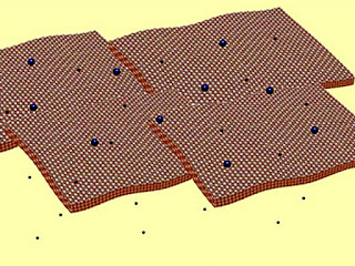
An illustration of rendered experimental data showing the polycrystalline copper surface and the differing graphene coverages. Graphene grows in a single layer on the (111) copper surface and in islands and multilayers elsewhere.

feature image Graphic by Joshua D. Wood
“However, this does not take into consideration the subtleties of growing graphene,” he said. “Understanding these subtleties is important for making high-quality, high-performance electronics.”

While graphene grown on copper tends to be better than graphene grown on other substrates, it remains riddled with defects and multi-layer sections, precluding high-performance applications. Researchers have speculated that the roughness of the copper surface may affect graphene growth, but the Illinois group found that the copper’s crystal structure is more important.

Copper foils are a patchwork of different crystal structures. As the methane falls onto the foil surface, the shapes of the copper crystals it encounters affect how well the carbon atoms form graphene.

Different crystal shapes are assigned index numbers. Using several advanced imaging techniques, the Illinois team found that patches of copper with higher index numbers tend to have lower-quality graphene growth. They also found that two common crystal structures, numbered (100) and (111), have the worst and the best growth, respectively. The (100) crystals have a cubic shape, with wide gaps between atoms. Meanwhile, (111) has a densely packed hexagonal structure.

“In the (100) configuration the carbon atoms are more likely to stick in the holes in the copper on the atomic level, and then they stack vertically rather than diffusing out and growing laterally,” Wood said. “The (111) surface is hexagonal, and graphene is also hexagonal. It’s not to say there’s a perfect match, but that there’s a preferred match between the surfaces.”

Researchers now are faced with balancing the cost of all (111) copper and the value of high-quality, defect-free graphene. It is possible to produce single-crystal copper, but it is difficult and prohibitively expensive.

The U. of I. team speculates that it may be possible to improve copper foil manufacturing so that it has a higher percentage of (111) crystals. Graphene grown on such foil would not be ideal, but may be “good enough” for most applications.

“The question is, how do you optimize it while still maintaining cost effectiveness for technological applications?” said Pop, a co-author of the paper. “As a community, we’re still writing the cookbook for graphene. We’re constantly refining our techniques, trying out new recipes. As with any technology in its infancy, we are still exploring what works and what doesn’t.”

Next, the researchers hope to use their methodology to study the growth of other two-dimensional materials, including insulators to improve graphene device performance. They also plan to follow up on their observations by growing graphene on single-crystal copper.

“There’s a lot of confusion in the graphene business right now,” Lyding said. “The fact that there is a clear observational difference between these different growth indices helps steer the research and will probably lead to more quantitative experiments as well as better modeling. This paper is funneling things in that direction.”

Lyding and Pop are affiliated with the Beckman Institute for Advanced Science and Technology at the U. of I. The Office of Naval Research, the Air Force Office of Scientific Research, and the Army Research Office supported this research.

Editor's note: To contact Joe Lyding, call 217-333-8370; email: lyding@illinois.edu.
To contact Eric Pop, call 217-244-2070; email: epop@illinois.edu.

Liz Ahlberg, Physical Sciences Editor | 217-244-1073; eahlberg@illinois.edu

NEWS BUREAU | PUBLIC AFFAIRS UNIVERSITY OF ILLINOIS 507 E. GREEN ST., SUITE 345, CHAMPAIGN, IL 61820 PH: 217-333-1085 FAX: 217-244-0161 E-MAIL: news@illinois.edu

Tuesday, October 25, 2011

Magnetic tunnel structures, thermoelectric voltages in nano-electronic junctions can be controlled

The heat which occurs in tiny computer processors might soon be no longer useless or even a problem. On the contrary: It could be used to switch these processors more easily or to store data more efficiently! These are two of the several potential applications made possible by a discovery made at the Physikalisch-Technische Bundesanstalt (PTB). This so-called "thermoelectronic voltage" may well be very interesting - mainly for the use of nano-junctions, i.e. small components based on magnetic tunnel structures. The results obtained by the researchers have been published in the current issue of the renowned specialised journal Physical Review Letters.

Today, magnetic tunnel structures already occur in various areas of information technology. They are used, for example, as magnetic storage cells in non-volatile magnetic memory chips (the so-called "MRAMs" - Magnetic Random Access Memories) or as highly sensitive magnetic sensors to read out the data stored on hard disks. The new effect discovered at PTB within the scope of a research collaboration with Bielefeld University and the Singulus company could, in the future, add a new application to the existing ones: monitoring and controlling thermoelectric voltages and currents in highly integrated electronic circuits.

Magnetic tunnel structures consist of two magnetic layers separated only by a thin insulation layer of approx. 1 nm - the so-called "tunnel barrier". The magnetic orientation of the two layers inside the tunnel structure has a great influence on its electrical properties: if the magnetic moments of the two layers are parallel to each other, the resistance is low; if, on the contrary, they are opposed to each other, the resistance is high. The change in the resistance when switching the magnetisation can amount to more than 100 %. It is therefore possible to control the electric current flowing through the magnetic tunnel structure efficiently by simply switching the magnetisation.

magnetic tunnel structure

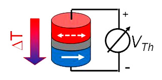
Schematic drawing of a magnetic tunnel structure with a tunnel magneto thermoelectric voltage (fig. by Schumacher/PTB)

A magnetic tunnel structure consists of two magnetic layers (red and blue) separated only by a thin insulation layer of approx. 1 nm (grey) - the so-called "tunnel barrier". If a temperature difference ?T is generated via the barrier, then the thermoelectric voltage VTh drops between the hot (red) layer and the cold (blue) layer. If the magnetic orientation, e.g. of the hot layer compared to that of the cold layer (arrows), is changed, this leads to a strong change in the measured thermoelectric voltage. (fig. by Schumacher/PTB)
The work carried out by the PTB researchers now shows that, besides the electric current, also the thermal current flowing through the tunnel structure can be influenced by switching the magnetisation. In their experiments, the scientists generated a temperature difference between the two magnetic layers and investigated the electric voltage (the so-called "thermoelectric voltage") generated hereby. It turned out that the thermoelectric voltage depends on the magnetic orientation of the two layers nearly as strongly as the electric resistance. By switching the magnetisation, it is therefore possible to control the thermoelectric voltage and, ultimately, also the thermal current flowing through the specimen.

In future, this new effect could be applied, for example, by using and converting the energy of waste heat occurring in integrated circuits in a targeted way. Furthermore, the discovery of this so-called "tunnel magneto thermoelectric voltage" is a milestone in the research field "spin calorics" - a field developing at a fast pace - which is currently promoted by the Deutsche Forschungsgemeinschaft (DFG) within the scope of a large-scale, 6-year priority programme.

Contact: Dr. Hans Werner Schumacher, Department 2.5 "Semiconductor Physics and Magnetism", Tel. +49 (0)531 592-2500, e-mail: hans.w.schumacher@ptb.de

Scientific publication: Liebing, N.; Serrano-Guisan, S.; Rott, K.; Reiss, G.; Langer, J.; Ocker, B.; Schumacher, H.W.: Tunneling magneto power in magnetic tunnel junction nanopillars. Phys. Rev. Lett. 107, 177201 (2011), prl.aps.org/abstract/PRL/v107/i17/e177201

Monday, October 24, 2011

If you've ever eaten from silverware or worn copper jewelry, you've been in a perfect storm in which nanoparticles were dropped into the environment

EUGENE, Ore. -- If you've ever eaten from silverware or worn copper jewelry, you've been in a perfect storm in which nanoparticles were dropped into the environment, say scientists at the University of Oregon.

Since the emergence of nanotechnology, researchers, regulators and the public have been concerned that the potential toxicity of nano-sized products might threaten human health by way of environmental exposure.

Now, with the help of high-powered transmission electron microscopes, chemists captured never-before-seen views of miniscule metal nanoparticles naturally being created by silver articles such as wire, jewelry and eating utensils in contact with other surfaces. It turns out, researchers say, nanoparticles have been in contact with humans for a long, long time.

The project involved researchers in the UO's Materials Science Institute and the Safer Nanomaterials and Nanomanufacturing Initiative (SNNI), in collaboration with UO technology spinoff Dune Sciences Inc. SNNI is an initiative of the Oregon Nanoscience and Microtechnologies Institute (ONAMI), a state signature research center dedicated to research, job growth and commercialization in the areas of nanoscale science and microtechnologies.

The research -- detailed in a paper placed online in advance of regular publication in the American Chemistry Society's journal ACS Nano -- focused on understanding the dynamic behavior of silver nanoparticles on surfaces when exposed to a variety of environmental conditions.

James E. Hutchison

James E. Hutchison
Using a new approach developed at UO that allows for the direct observation of microscopic changes in nanoparticles over time, researchers found that silver nanoparticles deposited on the surface of their SMART Grids electron microscope slides began to transform in size, shape and particle populations within a few hours, especially when exposed to humid air, water and light. Similar dynamic behavior and new nanoparticle formation was observed when the study was extended to look at macro-sized silver objects such as wire or jewelry.

"Our findings show that nanoparticle 'size' may not be static, especially when particles are on surfaces. For this reason, we believe that environmental health and safety concerns should not be defined -- or regulated -- based upon size," said James E. Hutchison, who holds the Lokey-Harrington Chair in Chemistry.

"In addition, the generation of nanoparticles from objects that humans have contacted for millennia suggests that humans have been exposed to these nanoparticles throughout time. Rather than raise concern, I think this suggests that we would have already linked exposure to these materials to health hazards if there were any."

Any potential federal regulatory policies, the research team concluded, should allow for the presence of background levels of nanoparticles and their dynamic behavior in the environment.

Because copper behaved similarly, the researchers theorize that their findings represent a general phenomenon for metals readily oxidized and reduced under certain environmental conditions. "These findings," they wrote, "challenge conventional thinking about nanoparticle reactivity and imply that the production of new nanoparticles is an intrinsic property of the material that is now strongly size dependent."

While not addressed directly, Hutchison said, the naturally occurring and spontaneous activity seen in the research suggests that exposure to toxic metal ions, for example, might not be reduced simply by using larger particles in the presence of living tissue or organisms.
###

Co-authors with Hutchison on the paper were Richard D. Glover, a doctoral student in Hutchison's laboratory, and John M. Miller, a research associate. Hutchison and Miller were co-founders of Dune Sciences Inc., a Eugene-based company that specializes in products and services geared toward the development and commercialization of nano-enabled products. Miller currently is the company's chief executive officer; Hutchison is chief science officer.

The electron microscopes used in this study are located at the Center for Advanced Materials Characterization in Oregon in the underground Lorry I. Lokey Laboratories at the UO. The U.S. Air Force Research Laboratory and W.M. Keck Foundation supported the research. Glover's participation also was funded by the National Science Foundation's STEM (science, technology, engineering, mathematics) Fellows in K-12 Education Program.

About the University of Oregon

The University of Oregon is among the 108 institutions chosen from 4,633 U.S. universities for top-tier designation of "Very High Research Activity" in the 2010 Carnegie Classification of Institutions of Higher Education. The UO also is one of two Pacific Northwest members of the Association of American Universities.

Contact: Jim Barlow, director of science and research communications, 541-346-3481, jebarlow@uoregon.edu Source: James E. Hutchison, Lokey-Harrington Chair in Chemistry, hutch@uoregon.edu. (NOTE: Hutchison currently is on sabbatical. To arrange an interview, contact him by email or through the media contact above)

Contact: Jim Barlow jebarlow@uoregon.edu 541-346-3481 University of Oregon

Sunday, October 23, 2011

Nanotechnology Measurement Championships VIDEO

Boston University contestant wins Inter-University Nanotechnology Measurement Championships

CAMBRIDGE, MA and CHRISTCHURCH, New Zealand - Dr. Meredith Mintzer a Postdoctoral Fellow at Boston University, was named the winner of the first Inter-University Nanotechnology Measurement Championships in Cambridge, Massachusetts last night. Contestants from Harvard University, MIT, Boston University and the University of Massachusetts raced each in the closely fought race to measure the particle concentration and size of a bimodal distribution of nanoparticles.

Around 120 attended the unique event hosted by nanotechnology instrument manufacturer Izon Science. Special guests included: Dr. Susan Windham-Bannister, Ph.D., President & CEO of the Massachusetts Life Sciences Center who presented the winner’s cup to Dr. Mintzer; Robert Coughlin, President and CEO of MassBio; and Ken Brown, Executive Director of the Massachusetts Office of International Trade and Investment.

Dr. Mintzer is from the laboratory of Prof. Mark Grinstaff, Boston University, Department of Biomedical Engineering. Dr. Mintzer uses Izon’s instruments in her research into drug delivery systems. The Grinstaff group pursues highly interdisciplinary research in the areas of biomedical engineering and macromolecular chemistry with the goal of elucidating the underlying fundamental chemistry and engineering principles of drug delivery systems.

Izon Science is the developer of the qNano and qViro nanotechnology instruments with unique size-tunable nanopores. The instruments offer significant improvements in accuracy and precision over previously available techniques and are helping to advance research in a number of fields including drug delivery, hematology, biomedical diagnostics, and vaccine development.

The Inter-University Nanoparticle Measurement Championships was held at an opening function for Izon Science’s new office and laboratory in Cambridge, MA which will serve as the company’s new US headquarters


The researchers - from Harvard University, MIT, Boston University & University of Massachusetts Lowell - who took part in the competition represent a broad range of research disciplines with particle characterization being the common theme.

VIDEO and TEXT CREDIT: nanoporenews

Hans van der Voorn Executive Chairman Izon Science Phone: + 64 21 463 399 Email: hans@izon.com Sandra Lukey Shine Group Phone: + 64 21 2262 858 Email: sandra@shinegroup.co.nz

About Izon Science:

Izon Science has developed the world’s first nanopore based measurement system available for general use. Izon’s instruments are used for precise measurement and analysis of individual particles across a wide range of scientific fields including bionanotechnology, nanomedicine, vaccinology, microbiology, biomedical research, environmental science, and particle based nanoscience. Izon originated in New Zealand and now sells its products in 23 countries. It has its European headquarters in Oxford, UK and US headquarters in Cambridge, MA Website: www.izon.com

Saturday, October 22, 2011

Transfer of fluoride anions between electrodes promises to enhance storage capacity reached by lithium-ion batteries by several factors

KIT researchers have developed a new concept for rechargeable batteries. Based on a fluoride shuttle - the transfer of fluoride anions between the electrodes – it promises to enhance the storage capacity reached by lithium-ion batteries by several factors. Operational safety is also increased, as it can be done without lithium. The fluoride-ion battery is presented for the first time in the “Journal of Materials Chemistry” by Dr. Maximilian Fichtner and Dr. Munnangi Anji Reddy.

Lithium-ion batteries are applied widely, but their storage capacity is limited. In the future, battery systems of enhanced energy density will be needed for mobile applications in particular. Such batteries can store more energy at reduced weight. For this reason, KIT researchers are also conducting research into alternative systems. A completely new concept for secondary batteries based on metal fluorides was developed by Dr. Maximilian Fichtner, Head of the Energy Storage Systems Group, and Dr. Munnangi Anji Reddy at the KIT Institute of Nanotechnology (INT).

Metal fluorides may be applied as conversion materials in lithium-ion batteries. They also allow for lithium-free batteries with a fluoride-containing electrolyte, a metal anode, and metal fluoride cathode, which reach a much better storage capacity and possess improved safety properties. Instead of the lithium cation, the fluoride anion takes over charge transfer. At the cathode and anode, a metal fluoride is formed or reduced. “As several electrons per metal atom can be transferred, this concept allows to reach extraordinarily high energy densities – up to ten times as high as those of conventional lithium-ion batteries,” explains Dr. Maximilian Fichtner.

fluoride-ion battery

Setup of the fluoride-ion battery: A fluoride-containing electrolyte separates the metal anode from the metal fluoride cathode. (Figure: KIT)
The KIT researchers are now working on the further development of material design and battery architecture in order to improve the initial capacity and cyclic stability of the fluoride-ion battery. Another challenge lies in the further development of the electrolyte: The solid electrolyte applied so far is suited for applications at elevated temperatures only. It is therefore aimed at finding a liquid electrolyte that is suited for use at room temperature.

M. Anji Reddy and M. Fichtner: Batteries based on fluoride shuttle. Journal of Materials Chemistry. 2011, Advance Article. DOI: 10.1039/C1JM13535J.

Karlsruhe Institute of Technology (KIT) is a public corporation according to the legislation of the state of Baden-Württemberg.

It fulfills the mission of a university and the mission of a national research center of the Helmholtz Association. KIT focuses on a knowledge triangle that links the tasks of research, teaching, and innovation.

Contact: Monika Landgraf presse@kit.edu 49-721-608-47414 Helmholtz Association of German Research Centres

Thursday, October 20, 2011

Slices of graphene single-atom form of carbon, in a solution form a nematic liquid crystal, particles are free-floating but aligned.

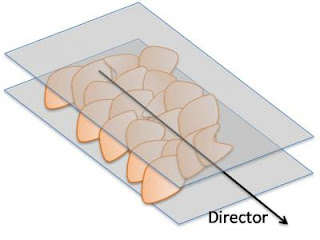
HOUSTON -- (Oct. 20, 2011) -- Giant flakes of graphene oxide in water aggregate like a stack of pancakes, but infinitely thinner, and in the process gain characteristics that materials scientists may find delicious.

A new paper by scientists at Rice University and the University of Colorado details how slices of graphene, the single-atom form of carbon, in a solution arrange themselves to form a nematic liquid crystal in which particles are free-floating but aligned.

That much was already known. The new twist is that if the flakes – in this case, graphene oxide – are big enough and concentrated enough, they retain their alignment as they form a gel. That gel is a handy precursor for manufacturing metamaterials or fibers with unique mechanical and electronic properties.

The team reported its discovery online this week in the Royal Society of Chemistry journal Soft Matter. Rice authors include Matteo Pasquali, a professor of chemical and biomolecular engineering and of chemistry; James Tour, the T.T. and W.F. Chao Chair in Chemistry as well as a professor of mechanical engineering and materials science and of computer science; postdoctoral research associate Dmitry Kosynkin; and graduate students Budhadipta Dan and Natnael Behabtu. Ivan Smalyukh, an assistant professor of physics at the University of Colorado at Boulder, led research for his group, in which Dan served as a visiting scientist.

"Graphene materials and fluid phases are a great research area," Pasquali said. "From the fundamental point of view, fluid phases comprising flakes are relatively unexplored, and certainly so when the flakes have important electronic properties.

Giant Graphene Oxide Flake

Caption: A single flake of graphene oxide roughly 40 microns wide, seen under an electron microscope, sits atop a copper support. Such "giant" flakes form into a gel-like liquid crystal in solution.

Credit: Rice University/University of Colorado at Boulder. Usage Restrictions: None.

Graphene oxide flakes

Caption: Graphene oxide flakes in a solution align themselves with a director, a dimensionless vector that represents the preferred orientation of particles in a liquid crystal.

Credit: Rice University/University of Colorado at Boulder. Usage Restrictions: None.
"From the application standpoint, graphene and graphene oxide can be important building blocks in such areas as flexible electronics and conductive and high-strength materials, and can serve as templates for ordering plasmonic structures," he said.

By "giant," the researchers referred to irregular flakes of graphene oxide up to 10,000 times as wide as they are high. (That's still impossibly small: on average, roughly 12 microns wide and less than a nanometer high.) Previous studies showed smaller bits of pristine graphene suspended in acid would form a liquid crystal and that graphene oxide would do likewise in other solutions, including water.

This time the team discovered that if the flakes are big enough and concentrated enough, the solution becomes semisolid. When they constrained the gel to a thin pipette and evaporated some of the water, the graphene oxide flakes got closer to each other and stacked up spontaneously, although imperfectly.

"The exciting part for me is the spontaneous ordering of graphene oxide into a liquid crystal, which nobody had observed before," said Behabtu, a member of Pasquali's lab. "It's still a liquid, but it's ordered. That's useful to make fibers, but it could also induce order on other particles like nanorods."

He said it would be a simple matter to heat the concentrated gel and extrude it into something like carbon fiber, with enhanced properties provided by "mix-ins."

Testing the possibilities, the researchers mixed gold microtriangles and glass microrods into the solution, and found both were effectively forced to line up with the pancaking flakes. Their inclusion also helped the team get visual confirmation of the flakes' orientation.

The process offers the possibility of the large-scale ordering and alignment of such plasmonic particles as gold, silver and palladium nanorods, important components in optoelectronic devices and metamaterials, they reported.

Behabtu added that heating the gel "crosslinks the flakes, and that's good for mechanical strength. You can even heat graphene oxide enough to reduce it, stripping out the oxygen and turning it back into graphite."

###

Co-authors of the paper are Angel Martinez and Julian Evans, graduate students of Smalyukh at the University of Colorado at Boulder.

The Institute for Complex Adaptive Matter, the Colorado Renewable and Sustainable Energy Initiative, the National Science Foundation, the Air Force Research Lab, the Air Force Office of Scientific Research, the Welch Foundation, the U.S. Army Corps of Engineers Environmental Quality and Installation Program and M-I Swaco supported the research.

Contact: Mike Williams mikewilliams@rice.edu 713-348-6728 Rice University

Wednesday, October 19, 2011

Microchip demonstrates concept of 'MRAM for biomolecules' using microfluidics and magnetic switches to trap and transport magnetic beads VIDEO

Researchers from the National Institute of Standards and Technology (NIST) and University of Colorado Boulder (CU) have developed a low-power microchip that uses a combination of microfluidics and magnetic switches to trap and transport magnetic beads. The novel transport chip may have applications in biotechnology and medical diagnostics.

A key innovation in the new chip is the use of magnetic switches like those in a computer random access memory. As described in a new paper,* the NIST/CU team used the chip to trap, release and transport magnetic beads that potentially could be used as transport vehicles for biomolecules such as DNA.

Conventional microfluidics systems use pumps and valves to move particles and liquids through channels. Magnetic particle transport microchips offer a new approach to microfluidics but generally require continuous power and in some cases cooling to avoid sample damage from excessive heating. The NIST/CU technology eliminates these drawbacks while offering the possibility for random access two-dimensional control and a memory that lasts even with the power off.

The demo chip features two adjacent lines of 12 thin-film magnet switches called spin valves, commonly used as magnetic sensors in read heads of high-density computer disk drives. In this case, however, the spin valves have been optimized for magnetic trapping. Pulses of electric current are used to switch individual spin valve magnets "on" to trap a bead, or "off" to release it, and thereby move the bead down a ladder formed by the two lines (see video clip). The beads start out suspended in salt water above the valves before being trapped in the array.


"It's a whole new way of thinking about microfluidics," says NIST physicist John Moreland. "The cool thing is it's a switchable permanent magnet—after it's turned on it requires no power. You beat heat by switching things quickly, so you only need power for less than a microsecond."

NIST researchers previously demonstrated that spin valves could be used to trap and rotate particles** and recently were awarded two patents related to the idea of a magnetic chip. ***

Magnetic tags are used in bioassays such as protein and DNA purification and cell breakdown and separation. The chip demonstration provides a conceptual foundation for a more complex magnetic random access memory (MRAM) for molecular and cellular manipulation. For example, programmable microfluidic MRAM chips might simultaneously control a large number of beads, and the attached molecules or cells, to assemble "smart" tags with specified properties, such as an affinity for a given protein at a specific position in the array. NIST is also interested in developing cellular and molecular tags for magnetic resonance imaging (MRI) studies in which individual cells, such as cancer cells or stem cells, would be tagged with a smart magnetic biomarker that can be tracked remotely in an MRI scanner, Moreland says. Automated spin valve chips might also be used in portable instruments for rapid medical diagnosis or DNA sequencing.

###

The lead author of the new paper, Wendy Altman, did the research at NIST as a CU graduate student working on her doctoral thesis. Another author, Bruce Han, was a CU student in NIST's Summer Undergraduate Research Fellowship (SURF) program.

* W.R. Altman, J. Moreland, S.E. Russek, B.W. Han and V. M. Bright. 2011. Controlled transport of superparamagnetic beads with spin-valves. Applied Physics Letters, Vol. 99, Issue 14, Oct. 3.

Contact: Laura Ost laura.ost@nist.gov 303-497-4880 National Institute of Standards and Technology (NIST)

Tuesday, October 18, 2011

Scientists build biological logic gates for digital computing devices out of bacteria and DNA

Scientists have successfully demonstrated that they can build some of the basic components for digital devices out of bacteria and DNA, which could pave the way for a new generation of biological computing devices, in research published today in the journal Nature Communications.

The researchers, from Imperial College London, have demonstrated that they can build logic gates, which are used for processing information in devices such as computers and microprocessors, out of harmless gut bacteria and DNA. These are the most advanced biological logic gates ever created by scientists.

Professor Richard Kitney, co-author of the paper from the Centre for Synthetic Biology and Innovation and the Department of Bioengineering at Imperial College London, says:

“Logic gates are the fundamental building blocks in silicon circuitry that our entire digital age is based on. Without them, we could not process digital information. Now that we have demonstrated that we can replicate these parts using bacteria and DNA, we hope that our work could lead to a new generation of biological processors, whose applications in information processing could be as important as their electronic equivalents.”

Although still a long way off, the team suggest that these biological logic gates could one day form the building blocks in microscopic biological computers. Devices may include sensors that swim inside arteries, detecting the build up of harmful plaque and rapidly delivering medications to the affected zone. Other applications may include sensors that detect and destroy cancer cells inside the body and pollution monitors that can be deployed in the environment, detecting and neutralising dangerous toxins such as arsenic.

biological logic gates

Professor Kitney and his team have created biological logic gates from bacteria and DNA.
The team say that the advantage of their biological logic gates over previous attempts is that they behave like their electronic counterparts. Previous research only proved that biological gates could be made. The new biological gates are also modular, which means that they can be fitted together to make different types of logic gates, paving the way for more complex biological processors to be built in the future.

In the new study, the researchers demonstrated how these biological logic gates worked. In one experiment, they showed how biological logic gates can replicate the way that electronic logic gates process information by either switching “on” or “off”.

The scientists constructed a type of logic gate called an “AND Gate” from bacteria called Escherichia coli (E.Coli), which is normally found in the lower intestine.

The team altered the E.Coli with modified DNA, which reprogrammed it to perform the same switching on and off process as its electronic equivalent when stimulated by chemicals.

The researchers were also able to demonstrate that the biological logic gates could be connected together to form more complex components in a similar way that electronic components are made. In another experiment, the researchers created a “NOT gate” and combined it with the AND gate to produce the more complex “NAND gate”.

The next stage of the research will see the team trying to develop more complex circuitry that comprises multiple logic gates. One of challenges faced by the team is finding a way to link multiple biological logic gates together similar to the way in which electronic logic gates are linked together to enable complex processing to be carried out.

For further information please contact:

Colin Smith Senior Research Media Officer Imperial College London Email: cd.smith@imperial.ac.uk Tel: +44(0)20 7594 6712. Out of hours duty press officer: +44(0)7803 886 248.

Notes to Editors:

1. " Engineering modular orthogonal genetic logic gates for robust digital-like synthetic biology ", 18 October 2011, Nature Communications journal.

The full listing of authors and their affiliations for this paper is as follows:

Baojan Wang (1), Richard Kitney (1), Nicolas Joly (2) (*) and Martin Buck (2)

(1) Centre for Synthetic Biology and Innovation and Department of Bioengineering, Imperial College, London SW7 2AZ, UK
(2) Division of Biology, Faculty of natural Sciences, Imperial College, London SW7 2AZ, UK
(*)Present address: institute Jacques monod, CNRS UMR 7592 , Universite Paris Diderot, Paris 75205, France.

2. About Imperial College London

Consistently rated amongst the world's best universities, Imperial College London is a science-based institution with a reputation for excellence in teaching and research that attracts 14,000 students and 6,000 staff of the highest international quality. Innovative research at the College explores the interface between science, medicine, engineering and business, delivering practical solutions that improve quality of life and the environment - underpinned by a dynamic enterprise culture.

Since its foundation in 1907, Imperial's contributions to society have included the discovery of penicillin, the development of holography and the foundations of fibre optics. This commitment to the application of research for the benefit of all continues today, with current focuses including interdisciplinary collaborations to improve global health, tackle climate change, develop sustainable sources of energy and address security challenges.

In 2007, Imperial College London and Imperial College Healthcare NHS Trust formed the UK's first Academic Health Science Centre. This unique partnership aims to improve the quality of life of patients and populations by taking new discoveries and translating them into new therapies as quickly as possible. Website: www.imperial.ac.uk

Sunday, October 16, 2011

Application calculates Casimir Effect forces and facilitates design of Microelectromechanical (MEMS) and Nanoelectromechanical (NEMS) systems

Dr. Martin Tajmar unveils a new nanotechnology software application that calculates Casimir Effect forces in nanotechnology design and facilitates the design of Microelectromechanical (MEMS) and future Nanoelectromechanical (NEMS) systems.

The CasimirSim software application assists scientists and engineers to design nanotechnology products by allowing them to model complex molecular geometries on the nanoscale, which should provide a greater understanding of how Casimir Effect forces impact nanotechnology.

Nanotechnology is definitely one of the leading technologies of the 21st century. It enables breakthroughs in physics, space applications, medicine, and materials sciences, to mention only a few examples. When scaling things down from our macroscopic everyday world to below one micrometer the nature of physics changes dramatically. The Casimir Effect is one example of quantum scale effects that play a role in the design of nanotechnology.

Quantum mechanical effects start to invalidate macroscopic models and govern the behavior of processes at the nano scale. CasimirSim has been developed for calculating Casimir Polder forces in arbitrary 3D geometries. CasimirSim is designed as a tool for scientists and engineers, therefore providing a wide variety of options for calculations. It shall open the doors for the analysis of expected Casimir forces in complex systems at design time, and help to approach new frontiers in nanotechnology.


TEXT and VIDEO CREDIT: americanantigravity

Saturday, October 15, 2011

Mobile electrons can be produced by the absorption of a single light particle in films of coupled quantum dots

Mobile electrons can be produced by the absorption of a single light particle in films of coupled quantum dots

Researchers of the Opto-electronic Materials section of TU Delft and Toyota Europe have demonstrated that several mobile electrons can be produced by the absorption of a single light particle in films of coupled quantum dots. These multiple electrons can be harvested in solar cells with increased efficiency. The researchers published their findings in the October issue of the scientific journal Nano Letters.

A way to increase the efficiency of cheap solar cells is the use of semiconductor nanoparticles, also called quantum dots. In theory, the efficiency of these cells can be increased to 44%. This is due to an interesting effect that efficiently happens in these nanoparticles: carrier multiplication. In the current solar cells, an absorbed light particle can only excite one electron, while in a quantum dot solar cell a light particle can excite several electrons. Multiplying the number of electrons results in the enhancement of current in solar cells, increasing the overall power conversion efficiency.

Carrier Multiplication

Several years ago it was demonstrated that carrier multiplication is more efficient in quantum dots than in traditional semiconductors. As a result, these quantum dots are currently heavily investigated worldwide for use in solar cells. A problem with using carrier multiplication is that the produced charges live only a very short time (around 0.00000000005 s) before they collide with each other and disappear via a decay process known as Auger recombination. The main current challenge is to proof that it is still possible to do something useful with them.

Mobile charges

mobile electrons multiplied in quantum dot films

Three for the price of one – mobile electrons multiplied in quantum dot films
The researchers from Delft have now demonstrated that even this very short time is long enough to separate the multiple electrons from each other. They prepared films of quantum dots in which the electrons can move so efficiently between the quantum dots that they become free and mobile before the time it takes to disappear via Auger recombination. In these films up to 3.5 free electrons are created per absorbed light particle. In this way, these electrons do not only survive, they are able to move freely through the material to be available for collection in a solar cell.

C. S. Suchand Sandeep,researcher with the section Opto-electronic Materials, Faculty of Applied Sciences, TU Delft. Phone: +31 (0) 15 278 3460, S.S.ChandramathiSukumaran@TUDelft.nl

Michiel Aerts, researcher with the section Opto-electronic Materials, Faculty of Applied Sciences, TU Delft. Phone: +31 (0) 15 278 3460, M.Aerts@TUDelft.nl

Sachin Kinge, Research & Development Toyota Europe. sachin.kinge@toyota-europe.com

InekeBoneschansker, science information officer at TU Delft. +31 (0) 15 278 8499, I.Boneschansker@TUDelft.nl

Thursday, October 13, 2011

Demonstrating a computer can analyze raw data from a biological system and derive mathematical equations that describe the way the system operates

First it was chess. Then it was Jeopardy. Now computers are at it again, but this time they are trying to automate the scientific process itself.

An interdisciplinary team of scientists at Vanderbilt University, Cornell University and CFD Research Corporation, Inc., has taken a major step toward this goal by demonstrating that a computer can analyze raw experimental data from a biological system and derive the basic mathematical equations that describe the way the system operates. According to the researchers, it is one of the most complex scientific modeling problems that a computer has solved completely from scratch.

The paper that describes this accomplishment is published in the October issue of the journal Physical Biology and is currently available online.

The work was a collaboration between John P. Wikswo, the Gordon A. Cain University Professor at Vanderbilt, Michael Schmidt and Hod Lipson at the Creative Machines Lab at Cornell University and Jerry Jenkins and Ravishankar Vallabhajosyula at CFDRC in Huntsville, Ala.

The "brains" of the system, which Wikswo has christened the Automated Biology Explorer (ABE), is a unique piece of software called Eureqa developed at Cornell and released in 2009. Schmidt and Lipson originally created Eureqa to design robots without going through the normal trial and error stage that is both slow and expensive. After it succeeded, they realized it could also be applied to solving science problems.

Robot biologist

Caption: This is a cartoon of robot biologist. Credit: Michael Smeltzer, Vanderbilt University. Usage Restrictions: None.
One of Eureqa's initial achievements was identifying the basic laws of motion by analyzing the motion of a double pendulum. What took Sir Isaac Newton years to discover, Eureqa did in a few hours when running on a personal computer.

In 2006, Wikswo heard Lipson lecture about his research. "I had a 'eureka moment' of my own when I realized the system Hod had developed could be used to solve biological problems and even control them," Wikswo said. So he started talking to Lipson immediately after the lecture and they began a collaboration to adapt Eureqa to analyze biological problems.

"Biology is the area where the gap between theory and data is growing the most rapidly," said Lipson. "So it is the area in greatest need of automation."

Software passes test: The biological system that the researchers used to test ABE is glycolysis, the primary process that produces energy in a living cell.

Specifically, they focused on the manner in which yeast cells control fluctuations in the chemical compounds produced by the process.

The researchers chose this specific system, called glycolytic oscillations, to perform a virtual test of the software because it is one of the most extensively studied biological control systems. Jenkins and Vallabhajosyula used one of the process' detailed mathematical models to generate a data set corresponding to the measurements a scientist would make under various conditions. To increase the realism of the test, the researchers salted the data with a 10 percent random error. When they fed the data into Eureqa, it derived a series of equations that were nearly identical to the known equations.

"What's really amazing is that it produced these equations a priori," said Vallabhajosyula. "The only thing the software knew in advance was addition, subtraction, multiplication and division."

Beyond Adam

The ability to generate mathematical equations from scratch is what sets ABE apart from Adam, the robot scientist developed by Ross King and his colleagues at the University of Wales at Aberystwyth. Adam runs yeast genetics experiments and made international headlines two years ago by making a novel scientific discovery without direct human input. King fed Adam with a model of yeast metabolism and a database of genes and proteins involved in metabolism in other species. He also linked the computer to a remote-controlled genetics laboratory. This allowed the computer to generate hypotheses, then design and conduct actual experiments to test them.

"It's a classic paper," Wikswo said.

In order to give ABE the ability to run experiments like Adam, Wikswo's group is currently developing "laboratory-on-a-chip" technology that can be controlled by Eureqa. This will allow ABE to design and perform a wide variety of basic biology experiments. Their initial effort is focused on developing a microfluidics device that can test cell metabolism.

"Generally, the way that scientists design experiments is to vary one factor at a time while keeping the other factors constant, but, in many cases, the most effective way to test a biological system may be to tweak a large number of different factors at the same time and see what happens. ABE will let us do that," Wikswo said.

###

The project was funded by grants from the National Science Foundation, National Institute on Drug Abuse, the Defense Threat Reduction Agency and the National Academies Keck Futures Initiative.

Contact: David F Salisbury david.salisbury@vanderbilt.edu 615-343-6803 Vanderbilt University

Wednesday, October 12, 2011

Molecular sieve could make production of gasoline, plastics and chemicals more cost effective and energy efficient

MINNEAPOLIS / ST. PAUL (10/12/2011) —A University of Minnesota team of researchers has overcome a major hurdle in the quest to design a specialized type of molecular sieve that could make the production of gasoline, plastics and various chemicals more cost effective and energy efficient. The breakthrough research, led by chemical engineering and materials science professor Michael Tsapatsis in the university's College of Science and Engineering, is published in the most recent issue of the journal Science.

After more than a decade of research, the team devised a means for developing free-standing, ultra-thin zeolite nanosheets that as thin films can speed up the filtration process and require less energy. The team has a provisional patent and hopes to commercialize the technology.

“In addition to research on new renewable fuels, chemicals and natural plastics, we also need to look at the production processes of these and other products we use now and try to find ways to save energy,” Tsapatsis said.

Separating mixed substances can demand considerable amounts of energy—currently estimated to be approximately 15 percent of the total energy consumption—part of which is wasted due to process inefficiencies. In days of abundant and inexpensive fuel, this was not a major consideration when designing industrial separation processes such as distillation for purifying gasoline and polymer precursors. But as energy prices rise and policies promote efficiency, the need for more energy-efficient alternatives has grown.

flaky crystal-type nanosheets

U of M researchers developed “carpets” of flaky crystal-type nanosheets that can be used to separate molecules as a sieve or as a membrane barrier in both research and industrial applications to save money and energy.
One promising option for more energy-efficient separations is high-resolution molecular separation with membranes. They are based on preferential adsorption and/or sieving of molecules with minute size and shape differences. Among the candidates for selective separation membranes, zeolite materials (crystals with molecular-sized pores) show particular promise.

While zeolites have been used as adsorbents and catalysts for several decades, there have been substantial challenges in processing zeolitic materials into extended sheets that remain intact. To enable energy-savings technology, scientists needed to develop cost-effective, reliable and scalable deposition methods for thin film zeolite formation.

The University of Minnesota team used sound waves in a specialized centrifuge process to develop “carpets” of flaky crystal-type nanosheets that are not only flat, but have just the right amount of thickness. The resulting product can be used to separate molecules as a sieve or as a membrane barrier in both research and industrial applications.

“We think this discovery holds great promise in commercial applications,” said Kumar Varoon, a University of Minnesota chemical engineering and materials science Ph.D. candidate and one of the primary authors of the paper published in Science. “This material has good coverage and is very thin. It could significantly reduce production costs in refineries and save energy.”

Members of the research team include Ph.D. candidates Kumar Varoon and Xueyi Zhang; postdoctoral fellows Bahman Elyassi and Cgun-Yi Sung; former students and Ph.D. graduates Damien Brewer, Sandeep Kumar, J. Alex Lee and Sudeep Maheshwari, graduate student Anudha Mittal; former undergraduate student Melissa Gettel; and faculty members Matteo Cococcioni, Lorraine Francis, Alon McCormick, K. Andre Mkhoyan and Michael Tsapatsis.

This research is being funded by the United States Department of Energy (including the Carbon Sequestration Program and the Catalysis Center for Energy Innovation – An Energy Frontier Center), the National Science Foundation and a variety of University of Minnesota partners.

Contacts: Rhonda Zurn, College of Science and Engineering, rzurn@umn.edu, (612) 626-7959 Preston Smith, University News Service, smith@umn.edu, (612) 625-0552

Monday, October 10, 2011

Creating the next generation of computer chips using Graphene’s ‘Big Mac’ wonder material

The world’s thinnest, strongest and most conductive material, discovered in 2004 at the University of Manchester by Professor Andre Geim and Professor Kostya Novoselov, has the potential to revolutionize material science.

Demonstrating the remarkable properties of graphene won the two scientists the Nobel Prize for Physics last year and Chancellor of the Exchequer George Osborne has just announced plans for a £50m graphene research hub to be set up.

Now, writing in the journal Nature Physics, the University of Manchester team have for the first time demonstrated how graphene inside electronic circuits will probably look like in the future.

By sandwiching two sheets of graphene with another two-dimensional material, boron nitride, the team created the graphene ‘Big Mac’ – a four-layered structure which could be the key to replacing the silicon chip in computers.

Because there are two layers of graphene completed surrounded by the boron nitride, this has allowed the researchers for the first time to observe how graphene behaves when unaffected by the environment.

Dr Leonid Ponomarenko, the leading author on the paper, said: “Creating the multilayer structure has allowed us to isolate graphene from negative influence of the environment and control graphene’s electronic properties in a way it was impossible before.

Dr Leonid Ponomarenko

Material discoveries: Dr Leonid Ponomarenko of Manchester University exploring graphene’s potential. Image by University of Manchester.
“So far people have never seen graphene as an insulator unless it has been purposefully damaged, but here high-quality graphene becomes an insulator for the first time.”

The two layers of boron nitrate are used not only to separate two graphene layers but also to see how graphene reacts when it is completely encapsulated by another material.

Professor Geim said: “We are constantly looking at new ways of demonstrating and improving the remarkable properties of graphene.”

“Leaving the new physics we report aside, technologically important is our demonstration that graphene encapsulated within boron nitride offers the best and most advanced platform for future graphene electronics.

It solves several nasty issues about graphene’s stability and quality that were hanging for long time as dark clouds over the future road for graphene electronics.

"We did this on a small scale but the experience shows that everything with graphene can be scaled up.”

“It could be only a matter of several months before we have encapsulated graphene transistors with characteristics better than previously demonstrated.”

Graphene is a novel two-dimensional material which can be seen as a monolayer of carbon atoms arranged in a hexagonal lattice.

Its remarkable properties could lead to bendy, touch screen phones and computers, lighter aircraft, wallpaper-thin HD TV sets and superfast internet connections, to name but a few.

The £50m Graphene Global Research and Technology Hub will be set up by the Government to commercialise graphene. Institutions will be able to bid for the money via the Engineering and Physical Sciences Research Council (EPSRC) – who funded work leading to the award of the Nobel prize long before the applications were realised.

Contact: Daniel Cochlin daniel.cochlin@manchester.ac.uk 44-161-275-8387 University of Manchester

Saturday, October 08, 2011

‘nanoparticles’, emitted from diesel engines could be affecting bees’ brains and damaging their inbuilt ‘sat-navs’

Scientists are investigating a possible link between tiny particles of pollution found in diesel fumes and the global collapse of honey bee colonies.

Professor Guy Poppy, an ecologist, Dr Tracey Newman, a neuroscientist, and their team from the University of Southampton, believe that minuscule particles, or ‘nanoparticles’, emitted from diesel engines could be affecting bees’ brains and damaging their inbuilt ‘sat-navs’. They believe this may stop worker bees finding their way back to the hive.

The team is also investigating the possibility that nanoparticles are one of a number of stress factors that could lead to a tipping point in bee health, which in turn could contribute to bee colony collapse.

“Diesel road-traffic is increasing in the UK and research from the US has shown that nanoparticles found in its fumes can be detrimental to the brains of animals when they are exposed to large doses. We want to find out if bees are affected in the same way – and answer the question of why bees aren’t finding their way back to the hive when they leave to find food,” explains Professor Poppy.

Bees are estimated to contribute billions to the world’s economy - £430 million a year to the UK alone - by pollinating crops, producing honey and supporting employment. Yet winter losses have led to the loss of tens of thousands of beehives year on year since 2007. The US has seen a 35 per cent unexplained drop in the number of hives in 2007, 2008 and 2009*.


Honey BeeExtensive research, including a recent United Nations Report, has so far not identified the cause of bee declines.

The team from the University of Southampton, including biologists, nanotechnology researchers and ecologists will test the behavioural and neurological changes in honey bees, after exposure to diesel nanoparticles.

Chemical ecologist Dr Robbie Girling, adds: “The diesel fumes may have a dual affect in that they may be mopping up flower smells in the air, making it harder for the bees to find their food sources.”

Recent research which has revealed more about the effects of nanoparticles has enabled scientists to investigate this possible link to bee colony collapse.

The three year study has been made possible by a Leverhulme Trust Research Project Grant of £156,000.

Colony Collapse Disorder Progress Report in PDF FORMAT

TEXT CREDIT: University of Southampton University Road Southampton SO17 1BJ Main switchboard: Tel. +44 (0)23 8059 5000 Fax +44 (0)23 8059 3131

Thursday, October 06, 2011

Developing new ways of computing that are beyond the scope of conventional silicon electronics

RIVERSIDE, Calif. – The University of California, Riverside has received a $1.85 million grant to develop a new way of computing that is beyond the scope of conventional silicon electronics.

The goal of the project is to speed up applications that process large amounts of data such as internet searching, data compression, and image recognition.

The money is awarded to UC Riverside under the nationwide "Nanoelectronics for 2020 and Beyond" competition sponsored by the National Science Foundation and the Nanoelectronics Research Initiative.

"Conventional silicon electronics will soon face its ultimate limits," said Roland Kawakami, a professor of physics and astronomy and the four-year grant's principal investigator. "Our approach is to utilize the spin degree of freedom to store and process information, which will allow the functions of logic and memory to be fully integrated into a single chip."

Spin is a fundamental characteristic property of electrons which causes them to behave as tiny magnets with a "north" and "south" pole. Electrons can occupy different spin states corresponding to different orientations for the magnetic poles. For spin-based computing, data is held in the spin state of the electron.

Kawakami explained that unlike more traditional approaches to improve electronics by building a better transistor, the current project has a far more transformative approach.

Roland Kawakami, University of California - Riverside

Caption: Roland Kawakami is a professor of physics and astronomy at UC Riverside.

Credit: UCR Strategic Communications. Usage Restrictions: None

Magnetologic Gate

Caption: The image shows a magnetologic gate, which consists of graphene contacted by several magnetic electrodes. Data is stored in the magnetic state of the electrodes, similar to the way data is stored in a magnetic hard drive. For the logic operations, electrons move through the graphene and use its spin state to compare the information held in the individual magnetic electrodes.

Credit: Kawakami lab, UC Riverside. Usage Restrictions: None.
"We are looking at a completely new architecture or framework for computing," he said. "This involves developing a new type of 'building-block' device known as a magnetologic gate that will serve as the engine for this technology – similar to the role of the transistor in conventional electronics. In addition, we will develop and design the circuits needed to utilize this device for specific functions, such as searching, sorting, and forecasting."

A magnetologic gate consists of graphene contacted by several magnetic electrodes. Data is stored in the magnetic state of the electrodes, similar to the way data is stored in a magnetic hard drive. For the logic operations, electrons move through the graphene and use its spin state to compare the information held in the individual magnetic electrodes.

The research project, which began Sept. 15, is a multicampus effort being led by UC Riverside. The research group of Jing Shi, a UCR professor of physics and astronomy, will work closely with Kawakami's research group on the project. They will be joined by Ilya Krivorotov at UC Irvine; Lu Sham at UC San Diego; Igor Zutic at SUNY Buffalo, NY; and Hanan Dery and Hui Wu at the University of Rochester, NY.

"Our team consists of experts in spintronics, magnetoresistive memory, theoretical physics, circuit design, and CMOS integration, a technology for constructing integrated circuits," said Kawakami, a member of UCR's Center for Nanoscale Science and Engineering.

The project is based on two major breakthroughs in nanoelectronics: The concept of spin-based computing using a magnetologic gate designed by Sham's group at UC San Diego in 2007; and the demonstration of tunneling spin injection and spin transport in graphene by Kawakami's group in 2010.

"Bringing these two results together, we find that graphene is the most promising material for developing magnetologic gates in terms of high speed, low energy usage, and operation at room temperature," Kawakami said.

Most of the experimental work will be done at UCR and UC Irvine. The circuit design and theory will be done at UC San Diego, the University of Rochester, and SUNY Buffalo.

###

The University of California, Riverside (www.ucr.edu) is a doctoral research university, a living laboratory for groundbreaking exploration of issues critical to Inland Southern California, the state and communities around the world. Reflecting California's diverse culture, UCR's enrollment has exceeded 20,500 students. The campus will open a medical school in 2013 and has reached the heart of the Coachella Valley by way of the UCR Palm Desert Graduate Center. The campus has an annual statewide economic impact of more than $1 billion.

A broadcast studio with fiber cable to the AT&T Hollywood hub is available for live or taped interviews. UCR also has ISDN for radio interviews. To learn more, call (951) UCR-NEWS.

Contact: Iqbal Pittalwala iqbal@ucr.edu 951-827-6050 University of California - Riverside

Tuesday, October 04, 2011

Ferroelectric transistor random access memory nonvolatile storage

WEST LAFAYETTE, Ind. - Researchers are developing a new type of computer memory that could be faster than the existing commercial memory and use far less power than flash memory devices.

The technology combines silicon nanowires with a "ferroelectric" polymer, a material that switches polarity when electric fields are applied, making possible a new type of ferroelectric transistor.

"It's in a very nascent stage," said doctoral student Saptarshi Das, who is working with Joerg Appenzeller, a professor of electrical and computer engineering and scientific director of nanoelectronics at Purdue's Birck Nanotechnology Center.

The ferroelectric transistor's changing polarity is read as 0 or 1, an operation needed for digital circuits to store information in binary code consisting of sequences of ones and zeroes.

The new technology is called FeTRAM, for ferroelectric transistor random access memory.

"We've developed the theory and done the experiment and also showed how it works in a circuit," he said.

Findings are detailed in a research paper that appeared this month in Nano Letters, published by the American Chemical Society.

The FeTRAM technology has nonvolatile storage, meaning it stays in memory after the computer is turned off. The devices have the potential to use 99 percent less energy than flash memory, a non-volatile computer storage chip and the predominant form of memory in the commercial market.

ferroelectric transistor random access memory

This diagram shows the layout for a new type of computer memory that could be faster than the existing commercial memory and use far less power than flash memory devices. The technology, called FeTRAM, combines silicon nanowires with a "ferroelectric" polymer, a material that switches polarity when electric fields are applied, making possible a new type of ferroelectric transistor. (Birck Nanotechnology Center, Purdue University)

"However, our present device consumes more power because it is still not properly scaled," Das said. "For future generations of FeTRAM technologies one of the main objectives will be to reduce the power dissipation. They might also be much faster than another form of computer memory called SRAM."

The FeTRAM technology fulfills the three basic functions of computer memory: to write information, read the information and hold it for a long period of time.

"You want to hold memory as long as possible, 10 to 20 years, and you should be able to read and write as many times as possible," Das said. "It should also be low power to keep your laptop from getting too hot. And it needs to scale, meaning you can pack many devices into a very small area. The use of silicon nanowires along with this ferroelectric polymer has been motivated by these requirements."

The new technology also is compatible with industry manufacturing processes for complementary metal oxide semiconductors, or CMOS, used to produce computer chips. It has the potential to replace conventional memory systems.

A patent application has been filed for the concept.

The FeTRAMs are similar to state-of-the-art ferroelectric random access memories, FeRAMs, which are in commercial use but represent a relatively small part of the overall semiconductor market. Both use ferroelectric material to store information in a nonvolatile fashion, but unlike FeRAMS, the new technology allows for nondestructive readout, meaning information can be read without losing it.

This nondestructive readout is possible by storing information using a ferroelectric transistor instead of a capacitor, which is used in conventional FeRAMs.

This work was supported by the Nanotechnology Research Initiative (NRI) through Purdue's Network for Computational Nanotechnology (NCN), which is supported by National Science Foundation.

Contact: Emil Venere: venere@purdjue.edu 765-494-4709 Purdue University

Monday, October 03, 2011

scientists combine tumor-targeting peptides and nanoparticles to eliminate glioblastoma in a previously untreatable mouse model

LA JOLLA, Calif., October 3, 2011 – Glioblastoma is one of the most aggressive forms of brain cancer. Rather than presenting as a well-defined tumor, glioblastoma will often infiltrate the surrounding brain tissue, making it extremely difficult to treat surgically or with chemotherapy or radiation. Likewise, several mouse models of glioblastoma have proven completely resistant to all treatment attempts. In a new study, a team led by scientists at Sanford-Burnham Medical Research Institute (Sanford-Burnham) and the Salk Institute for Biological Studies developed a method to combine a tumor-homing peptide, a cell-killing peptide, and a nanoparticle that both enhances tumor cell death and allows the researchers to image the tumors. When used to treat mice with glioblastoma, this new nanosystem eradicated most tumors in one model and significantly delayed tumor development in another. These findings were published the week of October 3 in the Proceedings of the National Academy of Sciences of the USA.

"This is a unique nanosystem for two reasons. First, linking the cell-killing peptide to nanoparticles made it possible for us to deliver it specifically to tumors, virtually eliminating the killer peptide's toxicity to normal tissues. Second, ordinarily researchers and clinicians are happy if they are able to deliver more drugs to a tumor than to normal tissues. We not only accomplished that, but were able to design our nanoparticles to deliver the killer peptide right where it acts—the mitochondria, the cell's energy-generating center," said Erkki Ruoslahti, M.D., Ph.D., senior author of the study and distinguished professor in both Sanford-Burnham's NCI-designated Cancer Center in La Jolla and the Center for Nanomedicine, a Sanford-Burnham collaboration with the University of California, Santa Barbara.

Erkki Ruoslahti, M.D., Ph.D.,
Caption: Erkki Ruoslahti, M.D., Ph.D., is a distinguished professor in Sanford-Burnham's NCI-designated Cancer Center and the Center for Nanomedicine, a Sanford-Burnham collaboration with the University of California, Santa Barbara.

Credit: Sanford-Burnham Medical Research Institute. Usage Restrictions: free to use only in association with this story, using provided caption and credit
The nanosystem developed in this study is made up of three elements. First, a nanoparticle acts as the carrier framework for an imaging agent and for two peptides (short proteins). One of these peptides guides the nanoparticle and its payload specifically to cancer cells and the blood vessels that feed them by binding cell surface markers that distinguish them from normal cells. This same peptide also drives the whole system inside these target cells, where the second peptide wreaks havoc on the mitochondria, triggering cellular suicide through a process known as apoptosis.

Together, these peptides and nanoparticles proved extremely effective at treating two different mouse models of glioblastoma. In the first model, treated mice survived significantly longer than untreated mice. In the second model, untreated mice survived for only eight to nine weeks.

In sharp contrast, treatment with this nanosystem cured all but one of ten mice. What's more, in addition to providing therapy, the nanoparticles could aid in diagnosing glioblastoma; they are made of iron oxide, which makes them—and therefore the tumors they target—visible by MRI, the same technique already used to diagnose many health conditions.

In a final twist, the researchers made the whole nanosystem even more effective by administering it to the mice in conjunction with a third peptide. Dr. Ruoslahti and his team previously showed that this peptide, known as iRGD, helps co-administered drugs penetrate deeply into tumor tissue. iRGD has been shown to substantially increase treatment efficacy of various drugs against human breast, prostate, and pancreatic cancers in mice, achieving the same therapeutic effect as a normal dose with one-third as much of the drug. Here, iRGD enhanced nanoparticle penetration and therapeutic efficacy.

"In this study, our patients were mice that developed glioblastomas with the same characteristics as observed in humans with the disease. We treated them systemically with the nanoparticles. Once the nanoparticles reached the tumors' blood vessels, they delivered their payload (a drug) directly to the cell's power producer, the mitochondria. By destroying the blood vessels and also some surrounding tumor cells, we were able to cure some mice and extend the lifespan of the rest," said Dinorah Friedmann-Morvinski, Ph.D., co-first author of the study and post-doctoral research associate in the laboratory of Inder Verma, Ph.D. at the Salk Institute.

###

The study was funded by: the National Cancer Institute and the National Institute of Allergy and Infectious Diseases, parts of the National Institutes of Health; the Leducq Foundation; the Merieux Foundation; Ipsen/Biomeasure; and the H.N. and Frances C. Berger Foundation. The full list of authors includes: Lilach Agemya, Center for Nanomedicine; Dinorah Friedmann-Morvinski, Salk Institute; Venkata Ramana Kotamraju, Center for Nanomedicine; Lise Roth, Center for Nanomedicine; Kazuki N. Sugahara, Sanford-Burnham; Olivier M. Girard, University of California, San Diego; Robert F. Mattrey, University of California, San Diego; Inder M. Verma, Salk Institute; and Erkki Ruoslahti, Sanford-Burnham's NCI-designated Cancer Center and Center for Nanomedicine at the University of California, Santa Barbara.

About Sanford-Burnham Medical Research Institute

Sanford-Burnham Medical Research Institute is dedicated to discovering the fundamental molecular causes of disease and devising the innovative therapies of tomorrow. Sanford-Burnham, with operations in California and Florida, is one of the fastest-growing research institutes in the country. The Institute ranks among the top independent research institutions nationally for NIH grant funding and among the top organizations worldwide for its research impact. From 1999 – 2009, Sanford-Burnham ranked #1 worldwide among all types of organizations in the fields of biology and biochemistry for the impact of its research publications, defined by citations per publication, according to the Institute for Scientific Information. According to government statistics, Sanford-Burnham ranks #2 nationally among all organizations in capital efficiency of generating patents, defined by the number of patents issued per grant dollars awarded.

Sanford-Burnham utilizes a unique, collaborative approach to medical research and has established major research programs in cancer, neurodegeneration, diabetes, and infectious, inflammatory, and childhood diseases. The Institute is especially known for its world-class capabilities in stem cell research and drug discovery technologies. Sanford-Burnham is a nonprofit public benefit corporation. For more information, please visit www.sanfordburnham.org.

Contact: Heather Buschman hbuschman@sanfordburnham.org 858-795-5343 Sanford-Burnham Medical Research Institute