Sunday, August 30, 2009

Natural compound stops retinopathy

Compound restores balance in eye with one treatment; affects macular degeneration and cancer too.

OKLAHOMA CITY – Researchers at the University of Oklahoma Health Sciences Center have found a way to use a natural compound to stop one of the leading causes of blindness in the United States. The research appears online this month in the journal Diabetes, a publication of the American Diabetes Association.

The discovery of the compound's function in inflammation and blood vessel formation related to eye disease means scientists can now develop new therapies –including eye drops – to stop diabetic retinopathy, a disease which affects as many as five million Americans with Type 1 and Type 2 diabetes.

Timothy Lyons and Jay Ma, University of Oklahoma

Caption: Dr. Timothy Lyons, left, director of the Harold Hamm Oklahoma Diabetes Center, stands with Dr. Jay Ma, principal investigator on the project and director of research for the diabetes center.

Credit: OU Medicine. Usage Restrictions: None.
"There is no good treatment for retinopathy, which is why we are so excited about this work. This opens an entirely new area for pharmaceutical companies to target," said Jay Ma, the principal investigator on the project and a research partner at the OU Health Sciences Center, Dean A. McGee Eye Institute and the Harold Hamm Oklahoma Diabetes Center.

Diabetic retinopathy is the most common diabetic eye disease and a leading cause of blindness in American adults.
It is caused by changes in blood vessels of the retina, the light-sensitive tissue at the back of the eye. In some people with diabetic retinopathy, blood vessels may swell and leak fluid. In other people, abnormal new blood vessels grow on the surface of the retina. Over time, diabetic retinopathy can get worse and cause some vision loss or blindness.

Oklahoma researchers found that this inflammation and leakage is caused by an imbalance of two systems in the eye. To restore balance, they delivered the new compound to cells using nanoparticle technology. The treatment in research models stopped the leakage, blocked inflammation and kept unwanted blood vessels from growing. Researchers are now testing the compound's uses for cancer and age-related macular degeneration. ###

Dr. Ma's research is funded by the American Diabetes Association and the National Institutes of Health.

Contact: Diane Clay diane-clay@ouhsc.edu 405-271-2323 University of Oklahoma

Friday, August 28, 2009

Nanotechnology may increase longevity of dental fillings

AUGUSTA, Ga. – Tooth-colored fillings may be more attractive than silver ones, but the bonds between the white filling and the tooth quickly age and degrade. A Medical College of Georgia researcher hopes a new nanotechnology technique will extend the fillings' longevity.

"Dentin adhesives bond well initially, but then the hybrid layer between the adhesive and the dentin begins to break down in as little as one year," says Dr. Franklin Tay, associate professor of endodontics in the MCG School of Dentistry. "When that happens, the restoration will eventually fail and come off the tooth."

Half of all tooth-colored restorations, which are made of composite resin, fail within 10 years, and about 60 percent of all operative dentistry involves replacing them, according to research in the Journal of the American Dental Association.

Dr. Franklin Tay, Medical College of Georgia

Caption: Dr. Franklin Tay, associate professor of endodontics in the Medical College of Georgia School of Dentistry, is studying a new nanotechnology technique to extend the longevity of tooth-colored fillings.

Credit: Medical College of Georgia. Usage Restrictions: None.
"Our adhesives are not as good as we thought they were, and that causes problems for the bonds," Dr. Tay says.

To make a bond, a dentist etches away some of the dentin's minerals with phosphoric acid to expose a network of collagen, known as the hybrid layer. Acid-etching is like priming a wall before it's painted; it prepares the tooth for application of an adhesive to the hybrid layer so that the resin can latch on to the collagen network. Unfortunately, the imperfect adhesives leave spaces inside the collagen that are not properly infiltrated with resin, leading to the bonds' failure.

Dr. Tay is trying to prevent the aging and degradation of resin-dentin bonding by feeding minerals back into the collagen network.
With a two year, $252,497 grant from the National Institute of Dental & Craniofacial Research, he will investigate guided tissue remineralization, a new nanotechnology process of growing extremely small, mineral-rich crystals and guiding them into the demineralized gaps between collagen fibers.

His idea came from examining how crystals form in nature. "Eggshells and abalone [sea snail] shells are very strong and intriguing," Dr. Tay says. "We're trying to mimic nature, and we're learning a lot from observing how small animals make their shells."

The crystals, called hydroxyapatite, bond when proteins and minerals interact. Dr. Tay will use calcium phosphate, a mineral that's the primary component of dentin, enamel and bone, and two protein analogs also found in dentin so he can mimic nature while controlling the size of each crystal.

Crystal size is the real challenge, Dr. Tay says. Most crystals are grown from one small crystal into a larger, homogeneous one that is far too big to penetrate the spaces within the collagen network. Instead, Dr. Tay will fit the crystal into the space it needs to fill. "When crystals are formed, they don't have a definite shape, so they are easily guided into the nooks and crannies of the collagen matrix," he says.

In theory, the crystals should lock the minerals into the hybrid layer and prevent it from degrading. If Dr. Tay's concept of guided tissue remineralization works, he will create a delivery system to apply the crystals to the hybrid layer after the acid-etching process.

"Instead of dentists replacing the teeth with failed bonds, we're hoping that using these crystals during the bond-making process will provide the strength to save the bonds," Dr. Tay says. "Our end goal is that this material will repair a cavity on its own so that dentists don't have to fill the tooth." ###

Contact: Paula Hinely phinely@mcg.edu 706-721-3646 Medical College of Georgia

Thursday, August 27, 2009

New statistical technique improves precision of nanotechnology data

Measuring nanomaterials

A new statistical analysis technique that identifies and removes systematic bias, noise and equipment-based artifacts from experimental data could lead to more precise and reliable measurement of nanomaterials and nanostructures likely to have future industrial applications.

Known as sequential profile adjustment by regression (SPAR), the technique could also reduce the amount of experimental data required to make conclusions, and help distinguish true nanoscale phenomena from experimental error. Beyond nanomaterials and nanostructures, the technique could also improve reliability and precision in nanoelectronics measurements – and in studies of certain larger-scale systems.

(left to right) are Zhong Lin Wang, V. Roshan Joseph, C.F. Jeff Wu and Xinwei Deng.

Caption: Georgia Tech researchers illustrate how their new statistical technique improves measurement of nanostructure properties by correcting data errors. Shown (left to right) are Zhong Lin Wang, V. Roshan Joseph, C.F. Jeff Wu and Xinwei Deng.

Credit: Georgia Tech Photo: Gary Meek. Usage Restrictions: None.
Accurate understanding of these properties is critical to the development of future high-volume industrial applications for nanomaterials and nanostructures because manufacturers will require consistency in their products.

"Our statistical model will be useful when the nanomaterials industry scales up from laboratory production because industrial users cannot afford to make a detailed study of every production run," said C. F. Jeff Wu, a professor in the Stewart School of Industrial and Systems Engineering at the Georgia Institute of Technology.
"The significant experimental errors can be filtered out automatically, which means this could be used in a manufacturing environment."

Sponsored by the National Science Foundation, the research was reported June 25, 2009 in the early edition of the journal Proceedings of the National Academy of Sciences. The paper is believed to be the first to describe the use of statistical techniques for quantitative analysis of data from nanomechanical measurements.

Nanotechnology researchers have long been troubled by the difficulty of measuring nanoscale properties and separating signals from noise and data artifacts. Data artifacts can be caused by such issues as the slippage of structures being studied, surface irregularities and inaccurate placement of the atomic force microscope tip onto samples.

In measuring the effects of extremely small forces acting on extremely small structures, signals of interest may be only two or three times stronger than experimental noise. That can make it difficult to draw conclusions, and potentially masks other interesting effects.

"In the past, we have really not known the statistical reliability of the data at this size scale," said Zhong Lin Wang, a Regents' professor in Georgia Tech's School of Materials Science and Engineering. "At the nanoscale, small errors are amplified. This new technique applies statistical theory to identify and analyze the data received from nanomechanics so we can be more confident of how reliable it is."

In developing the new technique, the researchers studied a data set measuring the deformation of zinc oxide nanobelts, research undertaken to determine the material's elastic modulus. Theoretically, applying force to a nanobelt with the tip of an atomic force microscope should produce consistent linear deformation, but the experimental data didn't always show that.

In some cases, less force appeared to create more deformation, and the deformation curve was not symmetrical. Wang's research team attempted to apply simple data-correction techniques, but was not satisfied with the results.

"The measurements they had done simply didn't match what was expected with the theoretical model," explained Wu, who holds a Coca-Cola chair in engineering statistics. "The curves should have been symmetric. To address this issue, we developed a new modeling technique that uses the data itself to filter out the mismatch step-by-step using the regression technique."

Ideally, researchers would search out and correct the experimental causes of these data errors, but because they occur at such small size scales, that would be difficult, noted V. Roshan Joseph, an associate professor in the Georgia Tech School of Industrial and Systems Engineering.

"Physics-based models are based on several assumptions that can go wrong in reality," he said. "We could try to identify all the sources of error and correct them, but that is very time-consuming. Statistical techniques can more easily correct the errors, so this process is more geared toward industrial use."

Beyond correcting the errors, the improved precision of the statistical technique could reduce the effort required to produce reliable experimental data on the properties of nanostructures. "With half of the experimental efforts, you can get about the same standard deviation as following the earlier method without the corrections," Wu said. "This translates into fewer time-consuming experiments to confirm the properties."

For the future, the research team – which includes Xinwei Deng and Wenjie Mai in addition to those already mentioned – plans to analyze the properties of nanowires, which are critical to the operation of a family of nanoscale electric generators being developed by Wang's research team. Correcting for data errors in these structures will require development of a separate model using the same SPAR techniques, Wu said.

Ultimately, SPAR may lead researchers to new fundamental explanations of the nanoscale world.

"One of the key issues today in nanotechnology is whether the existing physical theories can still be applied to explain the phenomena we are seeing," said Wang, who is also director of Georgia Tech's Center for Nanostructure Characterization and Fabrication. "We have tried to answer the question of whether we are truly observing new phenomena, or whether our errors are so large that we cannot see that the theory still works."

Wang plans to use the SPAR technique on future work, and to analyze past research for potential new findings. "What may have seemed like noise could actually be an important signal," he said. "This technique provides a truly new tool for data mining and analysis in nanotechnology." ###

Contact: John Toon jtoon@gatech.edu 404-894-6986 Georgia Institute of Technology Research News

Tuesday, August 25, 2009

University of Leicester researchers discover new fluorescent silicon nanoparticles

Research may ultimately track the uptake of drugs by the body's cells.

Researchers in the Department of Physics and Astronomy at the University of Leicester have developed a new synthesis method, which has led them to the discovery of fluorescent silicon nanoparticles and may ultimately help track the uptake of drugs by the body's cells.

Dr Klaus von Haeften explained: "A key advantage of the new method is the independent control of the nanoparticles' size and their surface properties. The method is extremely versatile and produces the fluorescent suspensions in one go. The findings may revolutionise the performance of electronic chips while satisfying the increasing demand for higher integration densities."

suspension of nanoparticles

Caption: This is a suspension of nanoparticles in a quarz-glass cell exposed to ultra violet light. Credit: Dr Klaus von Haeften, University of Leicester. Usage Restrictions: Must be used with credit.
The nanoparticles contain just a few hundred silicon atoms and their fluorescence were discovered after mixing them with water. This resulted in stability in fluorescence intensity over more than a three month period.

An interdisciplinary research project with the Department of Chemistry, led by Professor Chris Binns and Dr Glenn Burley, also incorporates this new method of synthesis. They are aiming to link nanoparticles to drugs involved in the diagnosis and treatment of cancer.
Professor of Nanoscience in the Department of Physics and Astonomy, Chris Binns said: "Nanotechnology, that is, the use of structures whose dimensions are on the nanometre scale, to build new materials and devices, appears to hold the key to future developments in a wide range of technologies, including materials, science, information technology and healthcare."
Dr von Haeften added: "The approach developed in Leicester could be a key step towards the production of a variety of biomedical sensors that could help track the uptake of drugs by cells."

The benign nature of silicon also makes the nanoparticles useful as fluorescent markers for tagging biologically sensitive materials. The light from a single nanoparticle can be readily detected.

The results of this work were published this week Applied Physics Letters journal by researchers Anthony Brewer and Klaus von Haeften. ###

Notes to Editors: For more information on this please contact Dr Klaus von Haeften, email kvh6@le.ac.uk tel 0116 252 3525 or Professor Chris Binns, email cb12@le.ac.uk, tel 0116 252 3585
suspension of nanoparticles

Caption: This is a suspension of nanoparticles in a quarz-glass cell exposed to ultra violet light. The nanoparticles emit deep-blue fluorescence. Credit: Dr Klaus von Haeften, University of Leicester. Usage Restrictions: Must be used with credit.
The research appears in: volume (94) of Appl. Phys. Lett. and the page number (261102)

Contact: Dr. Klaus von Haeften kvh6@le.ac.uk 01-162-523-525 University of Leicester

Sunday, August 23, 2009

MIT: A new approach to engineering for extreme environments VIDEO

Scientist creates model to design radiation-resistant materials.

CAMBRIDGE, Mass.--Composite materials such as fiberglass, which take on a mix of properties of their constituent compounds, have been around for decades. Now, an MIT materials scientist is taking composites to the nanoscale, where entirely new properties, not found in any of the original compounds, can emerge.

Michael Demkowicz, an assistant professor in MIT's Department of Materials Science and Engineering, is part of a team based at Los Alamos National Laboratory that recently received a federal Energy Frontier Research Centers grant to develop nanocomposite materials that can endure high temperatures, radiation and extreme mechanical loading. The ultimate goal is to use these materials in energy applications including nuclear power, fuel cells, solar energy and carbon sequestration.


Caption: In this video, radioactive particles bombarding the interface of a copper-niobium nanocomposite initially damage the material, but the damage is quickly contained.

Credit: Courtesy/Michael Demkowicz. Usage Restrictions: None.
"All sectors of energy production need materials that can withstand extreme conditions," says Demkowicz, whose model offers a new approach to designing nanocomposites with desirable traits.

There are many models that can take a proposed material structure and predict how it will behave. However, such trial-and-error approaches still require repeated cycles of manufacture and testing and are "an extremely costly and time-consuming way to come up with a new material," says Demkowicz.
His model tackles what materials scientists call "the inverse problem" — specifying a desired set of properties and then predicting which structures will deliver them — and could dramatically speed up the design process.

Radiation resistance

Demkowicz' first target is radiation-resistant materials, which could improve the efficiency and safety of nuclear power plants.

Normally, when metals are exposed to radiation, high-energy particles such as neutrons bump into individual atoms and knock them out of their crystal lattice. Like billiard balls, the displaced atoms bump into neighboring atoms, spreading damage in the form of "vacancies" (holes where an atom is missing), and "interstitials" (an extra atom squeezed in where it shouldn't be). Clusters of these defects can make the material brittle and weak.

The key to making nanocomposite materials resistant to radiation damage lies in the interfaces between layers of different materials. As the layers become thinner, the interfaces play a more dominant role in the material properties because the ratio of interface area to the material's total volume becomes larger. These interfaces give rise to novel properties not found in the original materials.

In some nanocomposites, vacancies and interstitials can get trapped at interfaces, where they have a higher likelihood of meeting. When that happens, the extra atom fills in the hole and the crystal structure is restored. Under some conditions it can appear as if there was no radiation damage remaining at all, says Demkowicz.

Materials resistant to radiation damage could eventually be used to line nuclear reactors, a function now performed by stainless steel. That could extend the lifetime of nuclear reactors and allow them to operate under higher radiation doses. Whereas current reactors consume only about one percent of their fuel, these improved reactors could burn a higher percentage of nuclear fuel and leave behind less waste.

Demkowicz has used his model, which is based on reproducing the mechanical interactions of groups of atoms, to design a nanocomposite with interfaces that resist radiation. The material, described in Physical Review Letters last year, is a mix of copper and the metal niobium and could not be used in a nuclear reactor because it absorbs neutrons and becomes radioactive. However, now that he knows copper-niobium is resistant to radiation damage, Demkowicz can use his modeling techniques to look for other materials that share that property.

Once a promising candidate is identified, it takes several years of testing before a new material can be approved for use in a nuclear reactor, so it will likely be at least a decade before any of his potential new materials can be used, says Demkowicz. ###

In addition to Demkowicz, several other MIT researchers are involved in the new Energy Frontier Research Centers. MIT will host two of the centers — the Center for Excitonics, led by Associate Professor Marc Baldo, and the Solid-State Solar-thermal Energy Conversion Center, led by Professor Gang Chen.

Contact: Elizabeth Thomson thomson@mit.edu 617-258-5402 Massachusetts Institute of Technology

Friday, August 21, 2009

Can a new implant coating technique create a new Six Million Dollar Man?

Tel Aviv University develops superior method for coating orthopaedic and dental implants.

Tel Aviv University researcher Prof. Noam Eliaz of the TAU School of Mechanical Engineering has developed an electrochemical process for coating metal implants which vastly improves their functionality, longevity and integration into the body.

The new process could vastly improve the lives of people who have undergone complicated total joint replacement surgeries so they can better walk, run and ultimately avoid rejection of the implant by their bodies.

"The surface chemistry, structure and morphology of our new coatings resemble biological material," explains Prof. Eliaz. "We've been able to enhance the integration of the coating with the mineralized tissue of the body, allowing more peoples' bodies to accept implants." His new coating resulted in a 33% decrease in the level of materials failure, or delamination, in these implants.

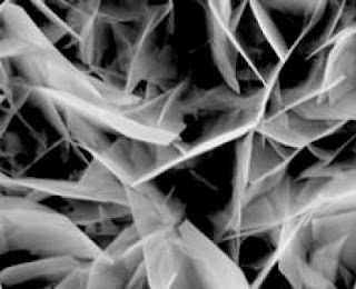
electrochemically-deposited crystals from a scanning electron microscope

Caption: This is an image of electrochemically-deposited crystals from a scanning electron microscope. Credit: AFTAU. Usage Restrictions: None.
Prof. Eliaz presented his findings to the 215th meeting of the Electrochemical Society in San Francisco in May 2009. In addition, a new 12-week implantation study, recently published in the journal Acta Biomaterialia, favorably compared the performance of the Tel Aviv University coatings to those of current commercial coatings.

Giving your joints an electrochemical bath

Today's surgeons can reconstruct joints in the human body using metal structures implanted to take the place of the natural joint.
In order to better integrate the new addition to the adjacent bone, implants are often coated with synthetic hydroxyapatite, which is similar to the main inorganic constituent of enamel, dentin and vertebrate bone. The properties of this coating are crucial to the function and life of the implant in the body.

Prof. Eliaz's advance is in the application technique of the coatings rather than the elements used in the coatings themselves. Instead of the traditional plasma-spraying technique, he and his team from the Tel Aviv University Materials and Nanotechnologies Program have developed a way to electrochemically deposit synthetic hydroxyapatite. In place of plasma-spraying the coating onto the metal, the metal implant is placed into a bath of electrolyte solution and an electric current is applied.
According to Prof. Eliaz, a good coating is crucial to the stable fixation of the implant in the surrounding bone. Since human bones naturally contain apatite, covering the implant with a synthetic version allows the body to register the implant as similar to a real bone. This ensures integration and fixation of the implant, and also prevents poisonous materials from leaking from the metal of the implant into the blood stream.Professor Noam Eliaz, Tel Aviv University

Caption: This is Professor Noam Eliaz of Tel Aviv University. Credit: AFTAU. Usage Restrictions: None
Could spur new bone growth

Prof. Eliaz has discovered that his method of coating circumvents the disadvantages of plasma- spraying. The electrochemical process allows synthetic hydroxyapatite to more closely mimic the real material. Examined under a microscope, it is virtually indistinguishable from the body's own material ― which helps the body accept a new implant.

The next-generation coating will include nano-particles to reinforce the coating. It will also have the potential to incorporate biological material or drugs during the process itself.

"We can incorporate biological materials" because the electrochemical process works at lower temperatures, says Prof. Eliaz. "The reinforcement of nanoparticles will improve the mechanical properties and may also improve the biological response. Drug incorporation may reduce the risk of post-surgery infection and even catalyze the growth of the bone." ###

American Friends of Tel Aviv University (www.aftau.org) supports Israel's leading and most comprehensive center of higher learning. In independent rankings, TAU's innovations and discoveries are cited more often by the global scientific community than all but 20 other universities worldwide.

Internationally recognized for the scope and groundbreaking nature of its research programs, Tel Aviv University consistently produces work with profound implications for the future.

Contact: George Hunka ghunka@aftau.org 212-742-9070 American Friends of Tel Aviv University

Wednesday, August 19, 2009

Stanford researchers find a quicker, cheaper way to sort isotopes

Isotopes, the atomic clues used to solve crimes, date ancient artifacts and identify chemicals.

Whether it's the summer grass that tickles your feet or the red Bordeaux smacking on your palette, nearly every part of the world around you carries special chemical markers. These markers, called isotopes, can tell scientists where the molecules that compose a substance are from, where they traveled, and what happened to them along the way. But doing these analyses has been complex and costly. Now, Stanford chemists have developed a new method to make isotopic analysis easier and less expensive.

"It's all done with smoke and mirrors," said chemist Richard Zare, giving a very literal description of the new method. The device he and his collaborators have created burns chemical samples into a gas, which then flows through a laser beam that is bouncing back and forth off a set of mirrors inside a special container.

Richard Zare, Stanford University

Richard Zare, Stanford University
The atoms of a particular element all have the same number of protons in their core, but may have differing numbers of neutrons. Carbon, for example, has six protons, but the number of neutrons in carbon atoms can vary from six to seven or eight. Each variation is an isotope of carbon.

Zare had the idea that it could be possible to distinguish different isotopes by the colors of light from the laser that they absorb when the original molecules are converted to smaller molecules through combustion.
"Think of them as being balls of different color," said Zare the Marguerite Blake Wilbur Professor in Natural Science and chair of the chemistry department. The tool can calculate the ratio of isotopes in a sample by simply "counting the colors and comparing them." This principle also makes the instrument more versatile than current mass spectrometers because Zare's device can analyze isotopes of different elements at the same time without being re-calibrated..

The equipment needed for the new method is smaller, cheaper, lighter and more portable than previous methods, and is easier to use. It has the potential to bring the power of isotopic analysis within easy reach of a host of researchers who have not had access to the expensive equipment that has been needed, Zare said. He and his collaborators report on their method in a paper scheduled to be published Monday, June 29, in the online early edition of the Proceedings of the National Academy of Sciences.

Isotopic analysis is used in a wide range of research, including geochemistry, medicine, and climatology. Until now, the analysis has been done using an isotope ratio mass spectrometer, which works by giving individual molecules an electric charge, then using a magnet to separate the isotopes by their mass—the more neutrons, the more mass. One machine can cost as much as a million dollars. In addition to being expensive and large, mass spectrometers now in use require specially trained technicians to operate them.

Zare's device, which employs what is called cavity ring-down spectroscopy, has potential applications in fields as varied as medicine, geology and winemaking, he said. "Some people are willing to pay a lot of money for wine," Zare said. "You allow me to measure the isotopes, I'll tell you whether you're paying your money for the real thing or not."

Because an element's isotopes are more plentiful in certain places than in others, the ratios of different isotopes within a larger mixture act like travel diaries – they can tell you the history of a mixture, whether it's from a different country, a particular part of the human body, or a previous time period. Determining the history of a mixture by measuring the ratios of its isotopes is known as isotopic analysis.

To illustrate, Zare explained that certain plants, such as corn, contain more carbon-13 than other plants. Because Americans tend to eat more corn than Europeans, isotopic analysis would detect more carbon-13 in the breath exhaled by an American than in the breath exhaled by a European, Zare said.

Doctors and pharmacologists can use isotopic analysis to measure the targeting precision of a specific drug by testing samples of urine and breath to see if the right organs have properly metabolized it. Also, climatologists can learn more about the ancient earth by studying carbon dioxide locked within cores of ice, Zare said.

Zare and his students worked together with researchers from Picarro, Inc., a start-up company he helped found, to create a prototype. They have successfully tested its performance by measuring carbon isotopes in different organic compounds such as methane, ethane, and propane. The bulky magnets that are the most expensive components of an isotope mass spectrometer are unnecessary in Zare's device, cutting costs while achieving an acceptable level of performance, according to Zare's team. Another advantage: the device can be used with minimal training.

Existing isotope ratio mass spectrometers can weigh as much as 1500 pounds and occupy the space of a large freezer case, such as those found an ice cream shop

Once the prototype is fully developed and commercialized, "It'll fit into the backseat of a car," Zare said. This portability can take isotopic analysis directly into the field, whether it's a doctor's office or a vineyard.

However, the team does see room for improvement.

The instrument's isotope ratio measurements are currently accurate within one to three parts per thousand, which is sufficient enough for the team to make a case for an alternative to isotope ratio mass spectrometry. However, this is still 10 to 30 times less accurate than isotope ratio mass spectrometers. The team emphasizes that their current results are preliminary and are only used to demonstrate the viability of their technique.

"My goal is to become better than and actually replace isotope ratio mass spectrometry," Zare said. He sees this as a possibility within the next 5 to 10 years. ###

Zare's co-authors on the paper include Stanford chemistry graduate student Douglas Kuramoto and Christa Haase, an undergraduate in chemistry at ETH, the Swiss Federal Institute of Technology, in Zurich, Switzerland, who worked in Zare's lab last summer. A grant from Picarro, Inc., a private gas analyzer manufacturer that Zare serves as a technical advisor and of which he is a founding member, supported the development of the current prototype. Other co-authors of the paper are Sze Tan, Eric Crosson, and Nabil Saad, researchers at Picarro, Inc.

Contact: Dan Stober dstober@stanford.edu 650-721-6965 Stanford University

Monday, August 17, 2009

Making nanoparticles in artificial cells

Two new construction manuals are now available for the world's smallest lamps. Based on these protocols, scientists from the Max Planck Institute of Colloids and Interfaces have tailor-made nanoparticles that can be used as position lights on cell proteins and, possibly in the future as well, as light sources for display screens or for optical information technology. The researchers produced cadmium sulphide particles in microscopically small membrane bubbles. Depending on which of the construction manuals they follow, the particles can be 4 or 50 nanometres in size. Because the membrane bubbles have the same size as living cells, the scientists' work also provides an indication as to how nanostructures could arise in nature. (Small, published online: June 8, 2009/DOI: 10.1002/smll.200900560)

Cells and microorganisms are absolute masters when it comes to working in the smallest possible dimensions. Like particularly efficient micro-factories, they produce particles and structures from inorganic material, for example pieces of chalk, that are only a few nanometres in size, that is, millionths of a millimetre. Cells have two different factors to thank for this capability. First, they have peptides, a biological tool at their disposal that may shape the chalk into a desired form. Second, the fact that they are very small themselves is convenient: the chalk particles cannot grow boundlessly - the end is reached when the calcium carbonate, the building block of chalk, runs out in the cell.

red fluorescent nanoparticles

Caption: Vesicles with different reactants have different fluorescent substances in their membranes (a). When the bubbles fuse, red fluorescent nanoparticles form (b). The particles can be seen as bright dots under the transmission electron microscope (c).

Credit: Image: Max Planck Institute of Colloids and Interfaces. Usage Restrictions: None.
"We used the fact that cells represent a closed reaction container as a model for the synthesis of nanoparticles," says Rumiana Dimova. Her group at the Max Planck Institute of Colloids and Interfaces studies membranes - the cell envelope.
The scientist and her colleagues form bubbles that are around 50 micrometres in size from lecithin membranes, which are similar to biological membranes. Like cells, membrane bubbles - or vesicles as scientists refer to them - also provide a closed reaction container. The scientists load the membrane bubbles with one of two reactants for the nanoparticles.

From this point, the researchers have developed two different sets of protocols. In one case, they produce bubbles loaded with one of the two reactants, sodium sulphide or cadmium chloride. The scientists then bring the bubbles with the different loads together and fuse two vesicles to form a bigger vesicle - this is done by subjecting the bubble cocktail to a short but very strong electrical pulse. The electric shock fuses the membranes of two adjacent bubbles.

In many cases, this results in the fusion of two bubbles containing different reactants. These then react to form cadmium sulphide, which is not water soluble and thus precipitates in the form of nanoparticles. "Because the reactants are only present to a limited extent in the fused bubbles, the particles only grow to a size of four nanometres," explains Rumiana Dimova. The scientists were able to track the entire process directly under the microscope because they had added different fluorescent molecules to the membranes of the differently loaded vesicles. The researchers were also able to see the nanoparticles forming as the particles shone like tiny lamps.

In the second process, the researchers only produce vesicles with one of the reactants. When the vesicles have formed, unlike in the first procedure, the researchers do not remove them from the production chamber. Instead, the bubbles remain attached to their substrate via small membrane channels, like balloons tied to strings, and stand in a solution that is the same as the one inside them. The researchers working with Rumiana Dimova then altered this situation: they substituted the solution with the first ingredient for the nanoparticles with a second component. This causes no change inside the vesicles at first. The second ingredient only creeps gradually between the substrate and membrane into the channel and to the vesicle. In the vesicle, where the other ingredient is already waiting, the nanoparticles grow again - this time to a size of 50 nanometres.

"With our method, we succeeded for the first time in producing particles with a certain diameter in vesicles whose size corresponds to that of cells," says Rumiana Dimova. Previously, biologists thought that cells depended on the help of peptides for the synthesis of nanoparticles. However, as Rumiana Dimova and her colleagues have discovered, it can also be done without them. ###

Original work:

Peng Yang, Reinhard Lipowsky, and Rumiana Dimova

Nanoparticle Formation in Giant Vesicles: Synthesis in Biomimetic Compartments

Small, published online, 8 June, 2009/DOI: 10.1002/smll.200900560

Contact: Dr. Rumiana Dimova Rumiana.Dimova@mpikg.mpg.de 49-331-567-9615 Max-Planck-Gesellschaft

Sunday, August 16, 2009

Implant bacteria, beware: Researchers create nano-sized assassins

PROVIDENCE, R.I. [Brown University] — Staphylococcus epidermidis is quite an opportunist. Commonly found on human skin, the bacteria pose little danger. But S. epidermidis is a leading cause of infections in hospitals. From catheters to prosthetics, the bacteria are known to hitch a ride on a range of medical devices implanted into patients.

Inside the body, the bacteria multiply on the implant's surface and then build a slimy, protective film to shield the colony from antibiotics. According to a study in the journal Clinical Infectious Diseases, up to 2.5 percent of hip and knee implants alone in the United States become infected, affecting thousands of patients, sometimes fatally.

More ominously, there is no effective antidote for infected implants. The only way to get rid of the bacteria is to remove the implant. "There is no [easy] solution," said Thomas Webster, a biomedical engineer at Brown University.

Erik Taylor, Brown University

Caption: Erik Taylor is a graduate student in engineering at Brown University. Credit: Brown University. Usage Restrictions: None.

Thomas Webster, Brown University

Caption: Thomas Webster is an associate professor in engineering and orthopedics at Brown University. Credit: Brown University. Usage Restrictions: None.
Now, Webster and Brown graduate student Erik Taylor have created a nano-sized headhunter that zeroes in on the implant, penetrates S. epidermidis's defensive wall and kills the bacteria. The finding, published in the International Journal of Nanomedicine, is the first time iron-oxide nanoparticles have been shown to eliminate a bacterial infection on an implanted prosthetic device.

In lab tests, Taylor, the lead author, and Webster, associate professor of engineering and orthopaedics, noted that up to 28 percent of the bacteria on an implant had been eliminated after 48 hours by injecting 10 micrograms of the nanoparticle agents. The same dosage repeated three times over six days destroyed essentially all the bacteria, the experiments showed.

The tests show "there will be a continual killing of the bacteria until the film is gone," said Webster, who is editor-in-chief of the peer-reviewed journal in which the paper appears.

A surprising added benefit, the scientists learned, is the nanoparticles' magnetic properties appear to promote natural bone cell growth on the implant's surface, although this observation needs to be tested further.

To carry out the study, the researchers created iron-oxide particles (they call them "superparamagnetic") with an average diameter of eight nanometers. They chose iron oxide because the metallic properties mean the particles can be guided by a magnetic field to the implant, while its journey can be tracked using a simple magnetic technique, such as magnetic resonance imaging (MRI).
Moreover, previous experiments showed that iron seemed to cause S. epidermidis to die, although researchers are unsure why. (Webster said it may be due to iron overload in the bacteria's cell.)
Once the nanoparticles arrive at the implant, they begin to penetrate the bacterial shield. The researchers are studying why this happens, but they believe it's due to magnetic horsepower. In the tests, the researchers positioned a magnet below the implant, producing a strong enough field to force the nanoparticles above to filter through the film and proceed to the implant, Webster explained.Iron-oxide nanoparticles

Caption: Iron-oxide nanoparticles developed at Brown University target an infected prosthesis, penetrate a bacterial film on the implant’s surface and thwart the colony by killing the bacteria. The nanoparticles also are believed to help natural bone cell growth.

Credit: Erik Taylor, Brown University Usage Restrictions: None.
The particles then penetrate the bacterial cells because of their super-small size. A micron-sized particle, a thousand times larger than a nanoparticle, would be too large to penetrate the bacterial cell wall. ###

The researchers plan to test the iron-oxide nanoparticles on other bacteria and then move on to evaluating the results on implants in animals. The research was funded by the private Hermann Foundation Inc. In addition, Taylor's tuition and stipend are funded through the National Science Foundation GK-12 program.

Contact: Richard Lewis Richard_Lewis@Brown.edu 401-863-3766 Brown University

Saturday, August 15, 2009

UGA researchers achieve breakthrough in effort to develop tiny biological fuel cells

University of Georgia researchers have developed a successful way to grow molecular wire brushes that conduct electrical charges, a first step in developing biological fuel cells that could power pacemakers, cochlear implants and prosthetic limbs. The journal Chemical Science calls the technique "a significant breakthrough for nanotechnology."

UGA chemist Jason Locklin and graduate students Nicholas Marshall and Kyle Sontag grew polymer brushes, made up of chains of thiophene and benzene, aromatic molecules sometimes used as solvents, attached to metal surfaces as ultra-thin films.

"The molecular wires are actually polymer chains that have been grown from a metal surface at very high density," said Locklin, who has a joint appointment in UGA's Franklin College of Arts and Science and on the Faculty of Engineering. "The structure of the film resembles a toothbrush, where the chains of conjugated polymers are like the bristles. We call these types of coatings polymer brushes. To get chains to pack tightly in extended conformations, they must be grown from the surface, a method we call the 'grafting from' approach."

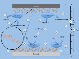
biofuel cell

This is an example of a biofuel cell in action. The red squiggly lines represent the polythiophene molecular wire brushes, by which direct electron transfer can occur between the enzyme and electrode (diagram by Kyle Sontag).
Using this approach, the scientists laid down a single layer of thiophene as the film's initial coating, then built up chains of thiophene or benzene using a controlled polymerization technique. Their research, funded by the Petroleum Research Foundation, was published in the June issue of the journal, Chemical Communications.

"The beauty of organic semiconductors is how their properties change, based on size and the number of repeating units," said Locklin, who is a member of UGA's Nanoscale Science and Engineering Center.
Thiophene itself is an insulator, said Locklin, "but by linking many thiophene molecules together in a controlled fashion, the polymers have conducting properties."

More importantly, he said, "this technique gives us the control to systematically vary polymer architecture, opening up the possibility for various uses in electronic devices such as sensors, transistors and diodes." The ultra-thin films are between 5 and 50 nanometers—too small to see, even under a high-powered optical microscope.

Locklin said it's difficult to harness a fuel source in the body, such as glucose, for use in biofuel cells that could replace the need for batteries in an implanted device. And while humans have enzymes in the body that do a good job of converting chemical energy into electrical energy, "they aren't very useful in this application because they have natural protective insulating layers that prevent good electron transport from active site to electrode," he said. "Hopefully our molecular wires will provide a better conduit for charges to flow."

While "flexible electronics" is a large and growing area of research, it's still in its infancy, Locklin said. "For example, we don't yet understand all of the fundamental physics involved in how electrical charges move through organic materials."

The next step for Locklin is to develop appropriate applications. For example, his polymer brush technique might be used in a range of devices that interface with living tissue, such as biochemical sensors, prosthetic limbs, pacemakers or bionic ears. "The film itself might be used in transistors—or in photovoltaic devices such as solar cells," said Locklin. ###

Contact: Jason Locklin jlocklin@chem.uga.edu 706-542-2359 University of Georgia

Thursday, August 13, 2009

Research explores interactions between nanomaterials, biological systems

Review article calls for measures to enable safe design of nanomaterials

The recent explosion in the development of nanomaterials with enhanced performance characteristics for use in commercial and medical applications has increased the likelihood of people coming into direct contact with these materials.

There are currently more than 800 products on the market — including clothes, skin lotions and cleaning products — claiming to have at least one nanocomponent, and therapeutic nanocarriers have been designed for targeted drug delivery inside the human body. Human exposure to nanomaterials, which are smaller than one one-thousandth the diameter of a human hair, raises some important questions, including whether these "nano-bio" interactions could have adverse health effects.

Andre Nel, M.B.Ch.B., Ph.D.

Andre Nel, M.B.Ch.B., Ph.D. Chief, Division of NanoMedicine, California. NanoSystems Institute. Professor, Medicine. Director, UC NanoToxicology Research Training Program. Member, California NanoSystems Institute, JCCC Signal Transduction and Therapeutics Program Area
Now, researchers at UCLA and the California NanoSystems Institute (CNSI), along with colleagues in academia and industry, have taken a proactive role in examining the current understanding of the nano-bio interface to identify the potential risks of engineered nanomaterials and to explore design methods that will lead to safer and more effective nanoparticles for use in a variety of treatments and products.

In a research review published in the July issue of the journal Nature Materials (and currently available online), the team provides a comprehensive overview of current knowledge on the physical and chemical properties of nanomaterials that allow them to undergo interactions with biological molecules and bioprocesses.
"What we have established here is a blueprint that will serve to educate the first generation of nanobiologists," said Dr. Andre Nel, leader of the team and chief of the division of nanomedicine at the David Geffen School of Medicine at UCLA and the California NanoSystems Institute.

Despite remarkable advances in nanoscience, relatively little is known about the intracellular activity and function of engineered nanomaterials, an area of study particularly important for the development of effective and safe nanoparticle drug-delivery systems. Much of the current knowledge derives from the study of tagged or labeled nanoparticles and their effects on cells after cellular uptake — without any detailed understanding of what these interactions may lead to, good or bad.

The review article examines the variety of ways in which nanomaterials interface with biological systems and presents a roadmap of the physical and chemical properties of the materials that could lead to potentially hazardous or advantageous interactions at the nano-bio interface. A better understanding of the biological impact, combined with appropriate stewardship, will allow for more informed decisions about design features for the safe use of nanotechnology.

In addition to Nel, the team included Tian Xia, a researcher in UCLA's nanomedicine division, UCLA associate professor of civil and environmental engineering Eric Hoek, Lutz Mädler of the University of Bremen, Darrell Velegol of Penn State University, Ponisseril Somasundaran of Columbia University, Fred Klessig of Pennsylvania Bio Systems, Vince Castranova of the National Institute for Occupational Safety and Health, and Mike Thompson of FEI Co.

"We are committed to ensuring that nanotechnology is introduced and implemented in a responsible and safe manner," said Nel, who also directs the Center for Environmental Implications of Nanotechnology, which is funded by the National Science Foundation and the Environmental Protection Agency and is headquartered at the CNSI.

"Based on our rapidly improving understanding of nano-bio interactions, we have done a thorough examination of the literature and our own research progress to identify measures that could be taken for safe design of nanomaterials," he said. "Not only will this improve the implementation and acceptance of this technology, but it will also provide the cornerstone of developing new and improved nanoscale therapeutic devices, such as drug-delivering nanoparticles."

The review article spotlighted several important research advancements:

* A classification of the interactions when nanomaterials contact and bind to biological systems will help scientists understand how man-made materials may react when exposed to cells, tissues and various life forms in different natural environmental contexts.

* When nanomaterials enter a biological fluid — for example, blood, plasma or interstitial fluid — the materials' surface may be coated with proteins. Understanding how these protein layers change the properties of the nanomaterials and the ways in which they interact in the body can provide valuable information on how to alter the protein coatings to allow for targeted delivery of nanomaterials to specific tissues, such as in cancer treatments.

* Physicochemical properties such as size, charge, shape and other characteristics could greatly affect the ability of nanomaterials to enter a cell; this could determine whether a material can be useful in nanomedicine applications or could cause harm if taken in by life forms in an ecosystem or food chain.

* Nanoparticles can elicit a wide range of intracellular responses, depending on their properties, concentrations and interactions with biological molecules. These properties and their relationships to cellular function can induce cellular damage or induce advantageous cellular responses, such as increased energy production and growth.

Based on the link between certain nanomaterial properties and potential toxic effects, the team asserts that scientists can reengineer specific nanomaterial properties that are hazardous while maintaining catalytically useful function for industrial use.

As an example of a safe design feature, some nanoparticles now receive a surface coating designed to improve safety by preventing bioreactivity. Nanoparticles in cosmetic formulations such as suntan lotions, for instance, may be coated with a water-repelling polymer to reduce direct contact with human skin. An extension of this principle uses polymers and detergents to decrease cellular uptake. However, there is the potential that when the coating wears off, the material may become hazardous. It is therefore important to consider improving the stability of coating substances. Coating nanoparticles with protective shells is also an effective means of preventing the breakup of materials that could release toxic substances upon dissolution.

"Instead of waiting for knowledge to unfold randomly, we can already begin to view the events at nano-bio interface as a discoverable scientific platform that can be used for setting up a deliberate inorganic-organic roadmap to new, better and safer products," Nel said. "What we can identify by understanding the rules that shape the nano-bio interface will have a massive impact on the ability to develop safe nanomaterials in the future."

###

The California NanoSystems Institute (CNSI) is an integrated research center operating jointly at UCLA and UC Santa Barbara whose mission is to foster interdisciplinary collaborations for discoveries in nanosystems and nanotechnology; train the next generation of scientists, educators and technology leaders; and facilitate partnerships with industry, fueling economic development and the social well-being of California, the United States and the world. The CNSI was established in 2000 with $100 million from the state of California and an additional $250 million in federal research grants and industry funding. At the institute, scientists in the areas of biology, chemistry, biochemistry, physics, mathematics, computational science and engineering are measuring, modifying and manipulating the building blocks of our world — atoms and molecules. These scientists benefit from an integrated laboratory culture enabling them to conduct dynamic research at the nanoscale, leading to significant breakthroughs in the areas of health, energy, the environment and information technology.

For more news, visit the UCLA Newsroom.

Contact: Jennifer Marcus jmarcus@cnsi.ucla.edu 310-267-4839 University of California - Los Angeles

Tuesday, August 11, 2009

Shape matters in the case of cobalt nanoparticles

Shape is turning out to be a particularly important feature of some commercially important nanoparticles—but in subtle ways. New studies* by scientists at the National Institute for Standards and Technology (NIST) show that changing the shape of cobalt nanoparticles from spherical to cubic can fundamentally change their behavior.

Building on a previous paper** that examined the properties of cobalt formed into spheres just a few nanometers in diameter, the new work explores what happens when the cobalt is synthesized instead as nanocubes. Nanoparticles of cobalt possess large magnetic moments—a measure of magnetic strength—and unique catalytic properties, and have potential applications in information storage, energy and medicine.

Cobalt Nanoparticles

Caption: These cubes of cobalt (left/top), measuring about 50 nanometers wide, are showing scientists that, on the nanoscale, a change in shape is a change in property. Unlike smaller spherical cobalt nanoparticles, nanocubes melt and fuse (right/bottom) when illuminated by a transmission electron microscope and possess different magnetic characteristics than the nanospheres as well.

Credit: NIST. Usage Restrictions: None.
One striking difference is the behavior of the two different particle types when external magnetic fields are applied and then removed. In the absence of a magnetic field, both the spherical and cubic nanoparticles spontaneously form chains—lining up as a string of microscopic magnets. Then, when placed in an external magnetic field, the individual chains bundle together in parallel lines to form thick columns aligned with the field. These induced columns, says NIST physicist Angela Hight Walker, imply that the external magnetic fields have a strong impact on the magnetic behavior of both nanoparticle shapes.
But their group interactions are somewhat different. As the strength of the external field is gradually reduced to zero, the magnetization of the spherical nanoparticles in the columns also decreases gradually. On the other hand, the magnetization of the cubic particles in the columns decreases in a much slower fashion until the particles rearrange their magnetic moments from linear chains into small circular groups, resulting in a sudden drop in their magnetization.

The team also showed that the cubes can be altered merely by observing with one of nanotechnology’s microscopes of choice. After a few minutes’ exposure to the illuminating beam of a transmission electron microscope, the nanocubes melt together, forming “nanowires” that are no longer separable as individual nanoparticles. The effect, not observed with the spheres, is surprising because the cubes average 50 nm across, much larger than the spheres’ 10 nm diameters. “You might expect the smaller objects to have a lower melting point,” Hight Walker says. “However, the sharp edges and corners in the nanocubes could be the locations to initiate melting.”

While Walker says that the melting effect could be a potential method for fabricating nanostructures, it also demands further attention. “This newfound effect demonstrates the need to characterize the physico-chemical properties of nanoparticles extremely well in order to pursue their applications in biology and medicine,” she says. ###

* G. Cheng, R.D. Shull and A.R. Hight Walker. Dipolar chains formed by chemically synthesized cobalt nanocubes. Journal of Magnetism and Magnetic Materials, May 11, 2009, Vol. 321, issue 10, pp. 1351—1355.

** G. Cheng, D. Romero, G.T. Fraser and A.R. Hight Walker. Magnetic-field-induced assemblies of cobalt nanoparticles. Langmuir, December 2005. See Oct. 20, 2007, Tech Beat article, “Magnetic Nanoparticles Assembled into Long Chains”.

Contact: Chad Boutin boutin@nist.gov 301-975-4261 National Institute of Standards and Technology (NIST)

Monday, August 10, 2009

Nanocrystals reveal activity within cells

Researchers at the U.S. Department of Energy's (DOE) Lawrence Berkeley National Laboratory have created bright, stable and bio-friendly nanocrystals that act as individual investigators of activity within a cell.

These ideal light emitting probes represent a significant step in scrutinizing the behaviors of proteins and other components in complex systems such as a living cell.

Labeling a given cellular component and tracking it through a typical biological environment is fraught with issues: the probe can randomly turn on and off, competes with light emitting from the cell, and often requires such intense laser excitation, it eventually destroys the probe, muddling anything you'd be interested in seeing.

Nanodetecttives

Caption: Berkeley Lab researchers have developed ideal single-molecule light emitting probes that represent a significant step in scrutinizing the behaviors of proteins and other components in complex systems such as a living cell.

Credit: courtesy of Jim Schuck, Molecular Foundry, Berkeley Lab. Usage Restrictions: None.

Nanocrystal Detective Team

Caption: Molecular Foundry post-doctoral researcher Shiwei Wu, staff scientist Jim Schuck, Facility Director Delia Milliron, staff scientist Bruce Cohen and post-doctoral researcher Gang Han demonstrate bright, stable and bio-friendly nanocrystal probes that act as individual investigators of their local environment.

Credit: Photo by Roy Kaltschmidt, Berkeley Lab Public Affirs. Usage Restrictions: None.
"The nanoparticles we've designed can be used to study biomolecules one at a time," said Bruce Cohen, a staff scientist in the Biological Nanostructures Facility at Berkeley Lab's nanoscience research center, the Molecular Foundry. "These single-molecule probes will allow us to track proteins in a cell or around its surface, and to look for changes in activity when we add drugs or other bioactive compounds."

Molecular Foundry post-doctoral researchers Shiwei Wu and Gang Han, led by Cohen, Imaging and Manipulation of Nanostructures staff scientist Jim Schuck and Inorganic Nanostructures Facility Director Delia Milliron, worked to develop nanocrystals containing rare earth elements that absorb low-energy infrared light and transform it into visible light through a series of energy transfers when they are struck by a continuous wave, near-infrared laser. Biological tissues are more transparent to near-infrared light, making these nanocrystals well suited for imaging living systems with minimal damage or light scatter.

"Rare earths have been known to show phosphorescent behavior, like how the old-style television screen glows green after you shut it off. These nanocrystals draw on this property, and are a million times more efficient than traditional dyes," said Schuck. "No probe with ideal single-molecule imaging properties had been identified to date—our results show a single nanocrystal is stable and bright enough that you can go out to lunch, come back, and the intensity remains constant."

To study how these probes might behave in a real biological system, the Molecular Foundry team incubated the nanocrystals with embryonic mouse fibroblasts, cells crucial to the development of connective tissue, allowing the nanocrystals to be taken up into the interior of the cell.
Live-cell imaging using the same near-infrared laser showed similarly strong luminescence from the nanocrystals within the mouse cell, without any measurable background signal.

"While these types of particles have existed in one form or another for some time, our discovery of the unprecedented 'single-molecule' properties these individual nanocrystals possess opens a wide range of applications that were previously inaccessible," Schuck adds. ###

"Non-blinking and photostable upconverted luminescence from single lanthanide-doped nanocrystals," by Shiwei Wu, Gang Han, Delia J. Milliron, Shaul Aloni, Virginia Altoe, Dmitri Talapin, Bruce E. Cohen and P. James Schuck, appears in Proceedings of the National Academy of Sciences and is available in Proceedings of the National Academy of Sciences online.

Work at the Molecular Foundry was supported by the Office of Basic Energy Sciences within the DOE Office of Science.

The Molecular Foundry is one of the five DOE Nanoscale Science Research Centers (NSRCs), premier national user facilities for interdisciplinary research at the nanoscale. Together the NSRCs comprise a suite of complementary facilities that provide researchers with state-of-the-art capabilities to fabricate, process, characterize and model nanoscale materials, and constitute the largest infrastructure investment of the National Nanotechnology Initiative. The NSRCs are located at DOE's Argonne, Brookhaven, Lawrence Berkeley, Oak Ridge and Sandia and Los Alamos National Laboratories. For more information about the DOE NSRCs, please visit nano.energy.gov.

Berkeley Lab is a U.S. Department of Energy national laboratory located in Berkeley, California. It conducts unclassified scientific research and is managed by the University of California. Visit our website at www.lbl.gov.

Contact: Aditi Risbud ASRisbud@lbl.gov 510-486-4861 DOE/Lawrence Berkeley National Laboratory

Saturday, August 08, 2009

Study gives clues to increasing X-rays' power

3-D, real-time X-ray images may be closer to reality

Three-dimensional, real-time X-ray images of patients could be closer to reality because of research recently completed by scientists at the University of Nebraska-Lincoln and a pair of Russian institutes.

In a paper to be published in an upcoming edition of Physical Review Letters, UNL Physics and Astronomy Professor Anthony Starace and his colleagues give scientists important clues into how to unleash coherent, high-powered X-rays.

"This could be a contributor to a number of innovations," Starace said.

Anthony F. Starace

Anthony F. Starace, George Holmes University Professor. Department of Physics & Astronomy. The University of Nebraska. 116 Brace Laboratory, Lincoln, NE 68588-0111

Office: B58 Behlen, E-mail: astarace1@unl.edu. Fax: (402) 472-2879, Phone: (402) 472-2795.
Starace's work focuses on a process called high-harmonic generation, or HHG. X-ray radiation can be created by focusing an optical laser into atoms of gaseous elements – usually low-electron types such as hydrogen, helium, or neon. HHG is the process that creates the energetic X-rays when the laser light interacts with those atoms' electrons, causing the electrons to vibrate rapidly and emit X-rays.

But the problem with HHG has been around almost as long as the onset of the method in 1988: The X-ray light produced by the atoms is very weak. In an effort to make the X-rays more powerful, scientists have attempted using higher-powered lasers on the electrons, but success has been limited.

"Using longer wavelength lasers is another way to increase the energy output of the atoms," Starace said. "The problem is, the intensity of the radiation (the atoms) produce drops very quickly."

Instead of focusing on low-electron atoms like hydrogen and helium, Starace's group applied HHG theory to heavier (and more rare) gaseous atoms having many electrons – elements such as xenon, argon and krypton.
They discovered that the process would unleash high-energy X-rays with relatively high intensity by using longer wavelength lasers (with wavelengths within certain atom-specific ranges) that happen to drive collective electron oscillations of the many-electron atoms.

"If you use these rare gases and shine a laser in on them, they'll emit X-Rays with an intensity that is much, much stronger (than with the simple atoms)," Starace said. "The atomic structure matters."

Starace said that unlocking the high-powered X-rays could lead one day, for example, to more powerful and precise X-ray machines. For instance, he said, heart doctors might conduct an exam by scanning a patient and creating a 3D hologram of his or her heart, beating in real time.

Nanoscientists, who study the control of matter on an atomic or molecular scale, also may benefit from this finding, Starace said. Someday, the high-intensity X-rays may be used to make 3D images of the microscopic structures with which nanoscientists work.

"With nanotechnology, miniaturization is the order of the day," he said. "But nanoscientists obviously could make use of a method to make the structures they're building and working with more easily visible." ###

The work is sponsored through funding by the National Science Foundation. Starace said NSF's sponsorship made the collaboration with his Russian colleagues – Mikhail V. Frolov, N.L. Manakov and T.S. Sarantseva of Voronezh State University, and M.Y. Emelin and M.Y. Ryabikin of the Russian Academy of Sciences – possible.

Frolov worked with Starace at UNL from 2002-2004 when he was a postdoctoral research associate in the Department of Physics and Astronomy. He has returned to Lincoln a number of times to collaborate with Starace on the HHG research. Frolov is a Ph.D. student of Manakov, with whom Starace has had a decade-long research collaboration that was initiated with support from NSF. Manakov also has an Adjunct Professor of Physics appointment in the UNL Department of Physics and Astronomy.

Contact: Steve Smith ssmith13@unl.edu 402-472-4226 University of Nebraska-Lincoln

Thursday, August 06, 2009

MIT slows concrete creep to a crawl

Work paves way for lightweight, vastly more durable infrastructure

CAMBRIDGE, Mass.--MIT civil engineers have for the first time identified what causes the most frequently used building material on earth — concrete — to gradually deform, decreasing its durability and shortening the lifespan of infrastructures such as bridges and nuclear waste containment vessels.

In a paper published in the Proceedings of the National Academy of Sciences (PNAS) online Early Edition the week of June 15, researchers say that concrete creep (the technical term for the time-dependent deformation that occurs in concrete when it is subjected to load) is caused by the rearrangement of particles at the nano-scale.

Franz-Josef Ulm

Professor Franz-Josef Ulm with the nanoindentation machine in his research lab. Photo / L. Barry Hetherington.

imprint left by a nanoindenter in a particle of cement

The image shows the imprint left by a nanoindenter in a particle of cement paste. The round blob at the top center is actually an extremely fine piece of dust on the surface. Photo / Chris Bobko
"Finally, we can explain how creep occurs," said Professor Franz-Josef Ulm, co-author of the PNAS paper. "We can't prevent creep from happening, but if we slow the rate at which it occurs, this will increase concrete's durability and prolong the life of the structures. Our research lays the foundation for rethinking concrete engineering from a nanoscopic perspective."

This research comes at a time when the American Society of Civil Engineers has assigned an aggregate grade of D to U.S. infrastructure, much of which is made of concrete. It likely will lead to concrete infrastructure capable of lasting hundreds of years rather than tens, which will bring enormous cost-savings and decreased concrete-related CO2 emissions. An estimated 5 to 8 percent of all human-generated atmospheric CO2 worldwide comes from the concrete industry.

Ulm, who has spent nearly two decades studying the mechanical behavior of concrete and its primary component, cement paste, has in the past several years focused on its nano-structure.
This led to his publication of a paper in 2007 that said the basic building block of cement paste at the nano-scale — calcium-silicate-hydrates, or C-S-H — is granular in nature. The paper explained that C-S-H naturally self-assembles at two structurally distinct but chemically similar phases when mixed with water, each with a fixed packing density close to one of the two maximum densities allowed by nature for spherical objects (64 percent for the lower density and 74 percent for high).

In the new research revealed in the PNAS paper, Ulm and co-author Matthieu Vandamme explain that concrete creep comes about when these nano-meter-sized C-S-H particles rearrange into altered densities: some looser and others more tightly packed.

They also explain that a third, more dense phase of C-S-H can be induced by carefully manipulating the cement mix with other minerals such as silica fumes, a waste material of the aluminum industry. These reacting fumes form additional smaller particles that fit into the spaces between the nano-granules of C-S-H, spaces that were formerly filled with water. This has the effect of increasing the density of C-S-H to up to 87 percent, which in turn greatly hinders the movement of the C-S-H granules over time.

"There is a search by industry to find an optimal method for creating such ultra-high-density materials based on packing considerations in confined spaces, a method that is also environmentally sustainable," said Ulm. "The addition of silica fumes is one known method in use for changing the density of concrete; we now know from the nanoscale packing why the addition of fumes reduces the creep of concrete. From a nanoscale perspective, other means now exist to achieve such highly packed, slow-creeping materials."

"The insight gained into the nanostructure puts concrete on equal footing with high-tech materials, whose microstructure can be nanoengineered to meet specific performance criteria: strength, durability and a reduced environmental footprint," said Vandamme, who earned a PhD from MIT's Department of Civil and Environmental Engineering in 2008 and is now on the faculty of the Ecole des Ponts ParisTech, Université Paris-Est.

In their PNAS paper, the researchers show experimentally that the rate of creep is logarithmic, which means slowing creep increases durability exponentially. They demonstrate mathematically that creep can be slowed by a rate of 2.6. That would have a truly remarkable effect on durability: a containment vessel for nuclear waste built to last 100 years with today's concrete could last up to 16,000 years if made with an ultra-high-density (UHD) concrete.

Ulm stressed that UHD concrete could alter structural designs, as well as have enormous environmental implications, because concrete is the most widely produced man-made material on earth: 20 billion tons per year worldwide with a 5 percent increase annually. More durable concrete means that less building material and less frequent renovations will be required.

"The thinner the structure, the more sensitive it is to creep, so up until now, we have been unable to build large-scale lightweight, durable concrete structures," said Ulm. "With this new understanding of concrete, we could produce filigree: light, elegant, strong structures that will require far less material."

Ulm and Vandamme achieved their research findings using a nano-indentation device, which allows them to poke and prod the C-S-H (or to use the terminology of civil engineering, apply load) and measure in minutes creep properties that are usually measured in year-long creep experiments at the macroscopic scale. ###

This work was funded in part by the Lafarge Group, a French building materials producer.

Contact: Elizabeth Thomson thomson@mit.edu 617-258-5402 Massachusetts Institute of Technology

Tuesday, August 04, 2009

Nanoparticle created to attack cardiovascular plaque

(Santa Barbara, Calif.) ––Scientists and engineers at UC Santa Barbara and the Burnham Institute for Medical Research have developed a nanoparticle that can attack plaque—a major cause of cardiovascular disease. The new development is described in a recent issue of the Proceedings of the National Academy of Sciences.

The treatment is promising for the eventual development of therapies for cardiovascular disease, which is blamed for one third of the deaths in the United States each year. Atherosclerosis, which was the focus of this study, is one of the leading causes of cardiovascular disease. In atherosclerosis, plaque builds up on the walls of arteries and can cause heart attack and stroke.

"The purpose of our grant is to develop targeted nanoparticles that specifically detect atherosclerotic plaques," said Erkki Ruoslahti, distinguished professor at the Burnham Institute for Medical Research at the University of California, Santa Barbara. "We now have at least one peptide, described in the paper, that is capable of directing nanoparticles to the plaques."

Multifunctional Micelle
Caption: This image shows a modular, multifunctional micelle created to target cardiovascular plaque and, when desired, carry a drug in the same particle directly to the plaque.

Credit: Peter Allen, UCSB College of Engineering. Usage Restrictions: None.
The nanoparticles in this study are lipid-based collections of molecules that form a sphere called a micelle. The micelle has a peptide, a piece of protein, on its surface, and that peptide binds to the surface of the plaque.

Co-author Matthew Tirrell, The Richard A. Auhll Professor and dean of UCSB's College of Engineering, specializes in lipid-based micelles. "This turned out to be a perfect fit with our targeting technology," said Ruoslahti.
To accomplish the research, the team induced atherosclerotic plaques in mice by keeping them on a high-fat diet. They then intravenously injected these mice with the micelles, which were allowed to circulate for three hours.

"One important element in what we did was to see if we could target not just plaques, but the plaques that are most vulnerable to rupture," said Ruoslahti. "It did seem that we were indeed preferentially targeting those places in the plaques that are prone to rupture."

The plaques tend to rupture at the "shoulder," where the plaque tissue meets the normal tissue. "That's also a place where the capsule on the plaque is the thinnest," said Ruoslahti. "So by those criteria, we seem to be targeting the right places."

Tirrell added:"We think that self-assembled micelles (of peptide amphiphiles) of the sort we have used in this work are the most versatile, flexible nanoparticles for delivering diagnostic and therapeutic biofunctionality in vivo. The ease with which small particles, with sufficiently long circulation times and carrying peptides that target and treat pathological tissue, can be constructed by self-assembly is an important advantage."

Ruoslahti said that UCSB's strength in the areas of materials, chemistry, and bioengineering facilitated this research. He noted that he and Tirrell have been close collaborators. ###

PNAS Paper Targeting atherosclerosis by using modular, multifunctional micelles:
www.pnas.org/content/

The work was funded by a grant from the National Heart, Lung and Blood Institute of the National Institutes of Health.

In addition to Ruoslahti and Tirrell, the article, "Targeting Atherosclerosis Using Modular, Multifunctional Micelles," was authored by David Peters of the Burnham Institute at UCSB and the Biomedical Sciences Graduate Group at UC San Diego; Mark Kastantin of UCSB's Department of Chemical Engineering; Venkata Ramana Kotamraju of the Burnham Institute at UCSB; Priya P. Karmali of the Cancer Research Center, Burnham Institute for Medical Research in La Jolla; and Kunal Gujraty of the Burnham Institute at UCSB.

Contact: Gail Gallessich gail.g@ia.ucsb.edu 805-893-7220 University of California - Santa Barbara

Sunday, August 02, 2009

Researchers solve 'bloodcurdling' mystery

Team uncovers the molecular basis for the regulation of blood clotting

CAMBRIDGE, Mass., June 4, 2009 – By applying cutting-edge techniques in single-molecule manipulation, researchers at Harvard University have uncovered a fundamental feedback mechanism that the body uses to regulate the clotting of blood. The finding, which could lead to a new physical, quantitative, and predictive model of how the body works to respond to injury, has implications for the treatment of bleeding disorders.
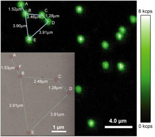
A team, co-led by Timothy A. Springer, Latham Family Professor of Pathology at Harvard Medical School and Children's Hospital Boston, and Wesley P. Wong, Rowland Junior Fellow and a Principal Investigator at the Rowland Institute at Harvard, reported its discovery about the molecular basis for the feedback loop responsible for hemostasis in the June 5th issue of Science.

Timothy A. Springer

The molecular construct was created in Timothy A. Springer's lab. File photograph by Kris Snibbe/Harvard News Office.
"The human body has an incredible ability to heal from life's scrapes and bruises," explains Wong. "A central aspect of this response to damage is the ability to bring bleeding to end, a process known as hemostasis. Yet regulating hemostasis is a complex balancing act."

Too much hemostatic activity can lead to an excess of blood clots, resulting in a potentially deadly condition known as thrombosis. If too little hemostatic activity occurs in the body, a person may bleed to death.

To achieve the proper balance, the body relies on a largely mechanical feedback system that relies on the miniscule forces applied by the circulation system on a molecular "force sensor" known as the A2 domain of the blood clotting protein von Willebrand factor (VWF).
By manipulating single molecules of this A2 domain, the researchers found that the A2 domain acts as a highly sensitive force sensor, responding to very weak tensile forces by unfolding, and losing much of its complex three-dimensional organization. This unfolding event allows the cutting of the molecule by an enzyme known as ADAMTS13.

"In the body, these cutting events decrease hemostatic potential and also enable blood clots to be trimmed in size. The system is so finely tuned that the A2 shear sensor is able to regulate the size of VWF within the blood stream, maintaining the optimal size for responding properly to traumas," says Wong.

To make the discovery, the team relied upon an "optical tweezers" system developed in Wong's lab. The tweezers are capable of applying miniscule forces to individual molecules while observing nanoscale changes in their length. Such manipulations enabled the researchers to characterize both the unfolding and refolding rates of single A2 molecules under force, as well as their interaction with the enzyme.

The molecular construct was created in Dr. Springer's lab, and consisted of an A2 domain connected to two DNA handles for manipulation. This elegant molecular system allowed the VWF "shear sensor" to be carefully studied and tested in isolation.

Ultimately, this work enhances the understanding of how the body is able to regulate the formation of blood clots, and is step towards a physical, quantitative, and predictive model of how the body responds to injury. It also gives insight into how bleeding disorders, such as type 2A von Willebrand disease, disrupt this regulation system, potentially leading to new avenues for treatment and diagnosis. ###

Wong and Springer's co-authors include Xiaohui Zhang, Kenneth Halvorsen, and Cheng-Zhong Zhang. The authors acknowledge the support of the National Institutes of Health, the American Heart Association, and the Rowland Junior Fellows program.

Contact: Michael Patrick Rutter mrutter@seas.harvard.edu 617-496-3815 Harvard University