Wednesday, December 31, 2008

Cold atoms could replace hot gallium in focused ion beams

Focused Ion Beams

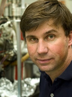
Caption: NIST researcher Jabez McClelland makes adjustments on the new magneto-optical trap ion source, capable of focusing beams of ions down to nanometer spots for use as a 'nano-scalpel' in advanced electronics processing.

Credit: Holmes, NIST. Usage Restrictions: None.
Scientists at the National Institute of Standards and Technology (NIST) have developed a radical new method of focusing a stream of ions into a point as small as one nanometer (one billionth of a meter).* Because of the versatility of their approach—it can be used with a wide range of ions tailored to the task at hand—it is expected to have broad application in nanotechnology both for carving smaller features on semiconductors than now are possible and for nondestructive imaging of nanoscale structures with finer resolution than currently possible with electron microscopes.
Researchers and manufacturers routinely use intense, focused beams of ions to carve nanometer-sized features into a wide variety of targets. In principle, ion beams also could produce better images of nanoscale surface features than conventional electron microscopy. But the current technology for both applications is problematic. In the most widely used method, a metal-coated needle generates a narrowly focused beam of gallium ions. The high energies needed to focus gallium for milling tasks end up burying small amounts in the sample, contaminating the material. And because gallium ions are so heavy (comparatively speaking), if used to collect images they inadvertently damage the sample, blasting away some of its surface while it is being observed. Researchers have tried using other types of ions but were unable to produce the brightness or intensity necessary for the ion beam to cut into most materials.

The NIST team took a completely different approach to generating a focused ion beam that opens up the possibility for use of non-contaminating elements. Instead of starting with a sharp metal point, they generate a small "cloud" of atoms and then combine magnetic fields with laser light to trap and cool these atoms to extremely low temperatures. Another laser is used to ionize the atoms, and the charged particles are accelerated through a small hole to create a small but energetic beam of ions. Researchers have named the groundbreaking device "MOTIS," for "Magneto-Optical Trap Ion Source." (For more on MOTs, see "Bon MOT: Innovative Atom Trap Catches Highly Magnetic Atoms," NIST Tech Beat Apr. 1, 2008.)

"Because the lasers cool the atoms to a very low temperature, they're not moving around in random directions very much. As a result, when we accelerate them the ions travel in a highly parallel beam, which is necessary for focusing them down to a very small spot," explains Jabez McClelland of the NIST Center for Nanoscale Science and Technology. The team was able to measure the tiny spread of the beam and show that it was indeed small enough to allow the beam to be focused to a spot size less than 1 nanometer. The initial demonstration used chromium atoms, establishing that other elements besides gallium can achieve the brightness and intensity to work as a focused ion beam "nano-scalpel." The same technique, says McClelland, can be used with a wide variety of other atoms, which could be selected for special tasks such as milling nanoscale features without introducing contaminants, or to enhance contrast for ion beam microscopy. ###

* J. L. Hanssen, S. B. Hill, J. Orloff and J. J. McClelland. Magneto-optical trap-based, high brightness ion source for use as a nanoscale probe. Nano Letters 8, 2844 (2008).

Contact: Mark Bello mark.bello@nist.gov 301-975-3776 National Institute of Standards and Technology (NIST)

Tuesday, December 30, 2008

New research expected to improve laser devices and make photovoltaics more efficient

Philippe Guyot-Sionnest

Philippe Guyot-Sionnest Professor Office: 929 E. 57th St., GCIS E 111, Chicago, IL 60637 Phone: (773)702-7461 Fax: (773)702-5863, Email:pgs@uchicago.edu
University of Chicago scientists have induced electrons in the nanocrystals of semiconductors to cool more slowly by forcing them into a smaller volume. This has the potential to improve satellite communications and the generation of solar power.

"Slowing down the cooling of these electrons—in this case, by more than 30 times—could lead to a better infrared laser source," said Philippe Guyot-Sionnest, Professor of Chemistry and Physics at the University of Chicago. "This, in turn, could be used to increase the bandwidth of communication satellites, allowing for faster connections."
Guyot-Sionnest is the principal investigator on the research project, which was described in a paper called "Slow Electron Cooling in Colloidal Quantum Dots," published Nov. 7 in Science.

The slow cooling of electrons in nanocrystals could lead to better, more efficient photovoltaic devices, he added. "This is because proposals to devise ways to extract the excess heat from these electrons as they cool are more likely to be realized—and to work—due to the fact that we now understand better what is going on with these nanocrystals."

Slower cooling of electrons in nanocrystals was first theorized in 1990, but no one has been able to observe this effect.

Slow electron cooling in nanocrystals occurs because forcing the electrons into a smaller volume leads them to oscillate between their alternate extremes within a very short period of time. (This is analogous to the way shorter strings on musical instruments produce higher pitches.) The electrons in the nanocrystals used in this experiment oscillated so fast that it became difficult for them to drag along the more sluggish vibrations of the nuclei. As a result, the energy stayed with the electrons for a longer period of time.

The slower cooling effect was difficult to induce and observe because several different mechanisms for energy loss interfered with the process. By eliminating these other mechanisms, the researchers were able to induce and observe slower electron cooling in nanocrystals. ###

Anshu Pandey, a graduate student in Chemistry at the University of Chicago, did the experiments described in the Science paper, which he co-authored.

Contact: Greg Borzo gregborzo@uchicago.edu 773-702-8366 University of Chicago

Monday, December 29, 2008

Survey highlights support for nanotech in health fields but disapproval elsewhere

Dr. Michael Cobb

Dr. Michael Cobb
A landmark national survey on the use of nanotechnology for "human enhancement" shows widespread public support for applications of the new technology related to improving human health. However, the survey also shows broad disapproval for nanotech human enhancement research in areas without health benefits. A team of researchers at North Carolina State University and Arizona State University (ASU) conducted the study, which could influence the direction of future nanotechnology research efforts.
The "Public Awareness of Nanotechnology Study" is the first nationally representative survey to examine public opinion on the use of nanotechnology for human enhancement. The survey found significant support for enhancements that promise to improve human health. For example, 88 percent of participants were in favor of research for a video-to-brain link that would amount to artificial eyesight for the blind. However, there was little support for non-health research endeavors. For example, only 30 percent of participants approved of research into implants that could improve performance of soldiers on the battlefield.

Nanotechnology is generally defined as technology that uses substances having a size of 100 nanometers or less (tens of thousands of times smaller than the width of a human hair), and is expected to have widespread uses in medicine, consumer products and industrial processes. Human enhancement is a sweeping term that applies to the use of such technologies to alter human capabilities.

NC State's Dr. Michael Cobb, one of the leaders of the study, says the survey's findings are important because "what the public wants could drive the direction of future research." Cobb, an associate professor of political science, explains, "The public should have input into where the government invests its research funding." Dr. Clark Miller, an associate professor of political science at ASU and another leader of the survey, adds, "One of the most important findings is the difference in support for different applications of human enhancement. Research and public policies will need to reflect this differentiated view, recognizing that there are some applications the public supports and some that the public is quite skeptical of."

While the survey shows strong public support for research into nanotechnology applications in the health field, those findings are tempered by a similar concern from the public about the scope of that research. The study found that 55 percent of participants felt that researchers should "avoid playing God with new technologies." Similarly, the public expressed little confidence in the government and mass media to inform people about potential risks from new technologies. Rather, participants said they had the greatest confidence in university scientists and environmental groups to protect the public. ###

Leaders of the study were NC State's Cobb, ASU's Miller, Sean Hays, doctoral student in political science at ASU, and Dr. David Guston, professor of political science and director of the Center for Nanotechnology in Society at Arizona State University (CNS). The study was funded by CNS under a cooperative agreement from the National Science Foundation to conduct research, training and outreach on the societal aspects of nanotechnology. The study's findings complement earlier findings of the CNS National Citizens' Technology Forum, organized by Cobb and NC State researcher Dr. Patrick Hamlett in April 2008.

The survey was conducted between July and October of 2008. The survey included 556 participants, had a 28 percent response rate, and has a margin of error of plus or minus 4.1 percent.

Contact: Matt Shipman matt_shipman@ncsu.edu 919-515-6386 North Carolina State University

Tags: or and

Sunday, December 28, 2008

Findings suggest nanowires ideal for electronics manufacturing VIDEO

nanowires form from gold nanoparticles exposed to a gas containing silicon

This image was taken from an animation depicting how tiny silicon "nanowires" form from gold nanoparticles exposed to a gas containing silicon. Researchers at Purdue and IBM's Thomas J. Watson Research Center used an instrument called a transmission electron microscope to watch how nanowires made of silicon "nucleate," or begin to form before growing into wires. Findings showed that the process is likely highly repeatable, meaning that the wires probably can be manufactured for future computers and electronics. (Image courtesy of Seyet LLC)
WEST LAFAYETTE, Ind. - Researchers have discovered that tiny structures called silicon nanowires might be ideal for manufacturing in future computers and consumer electronics because they form the same way every time.

The researchers use an instrument called a transmission electron microscope to watch how nanowires made of silicon "nucleate," or begin to form, before growing into wires, said Eric Stach, an assistant professor of materials engineering at Purdue University.

The work is based at IBM's Thomas J. Watson Research Center in Yorktown Heights, N.Y., and at Purdue's Birck Nanotechnology Center in the university's Discovery Park.
The nucleation process can be likened to the beginning of ice forming in a pool of water placed in a freezer. The liquid undergoes a "phase transition," changing from the liquid to the solid phase.

"What's unusual about this work is that we are looking at these things on an extremely small scale," Stach said. "The three major findings are that you can see that the nucleation process on this small scale is highly repeatable, that you can measure and predict when it's going to occur, and that those two facts together give you a sense that you could confidently design systems to manufacture these nanowires for electronics."

It was the first time researchers had made such precise measurements of the nucleation process in nanowires, he said.
Findings will be detailed in a research paper appearing Friday (Nov. 14) in the journal Science. The paper was written by Purdue doctoral student Bong Joong Kim and Stach and IBM materials scientists Frances Ross, Jerry Tersoff, Suneel Kodambaka and Mark Reuter from the physical sciences department at the Watson Research Center.

Real time observations of nanowire nucleation
The silicon nanowires begin forming from tiny gold nanoparticles ranging in size from 10 to 40 nanometers, or billionths of a meter. By comparison, a human red blood cell is more than 100 times larger than the gold particles.

The gold particles are placed in the microscope's vacuum chamber and then exposed to a gas containing silicon, and the particles act as a catalyst to liberate silicon from the gas to form into solid wires. The particles are heated to about 600 degrees Celsius, or more than 1,100 degrees Fahrenheit, causing them to melt as they become "supersaturated" with silicon from the gas. With increasing exposure, the liquid gold eventually contains too much silicon and the silicon precipitates as a solid, causing the nanowire to begin forming.

"We found that there is a single nucleation event in each little droplet and that all of the nucleation events occur in a very controllable fashion," Stach said. "The implication is that if you are trying to create electronic devices based on these technologies, you could actually predict when things are going to start their crystal growth process. You can see that it's going to happen the same way every time, and thus that there is some potential for doing things in a repeatable fashion in electronics manufacturing."

The research is funded by the National Science Foundation through the NSF's Electronic Materials Division.

Although the researchers studied silicon, the same findings could be applied to manufacturing nanowires made of other semiconducting materials. The electron microscope is the only instrument capable of observing the nanowire nucleation process, which would have to be a thousand times larger to be seen with a light microscope, Stach said.

Nanowires might enable engineers to solve a problem threatening to derail the electronics industry. New technologies will be needed for industry to keep pace with Moore's law, an unofficial rule stating that the number of transistors on a computer chip doubles about every 18 months, resulting in rapid progress in computers and telecommunications. Doubling the number of devices that can fit on a computer chip translates into a similar increase in performance. However, it is becoming increasingly difficult to continue shrinking electronic devices made of conventional silicon-based semiconductors.

"In something like five to, at most, 10 years, silicon transistor dimensions will have been scaled to their limit," Stach said.

Transistors made of nanowires represent one potential way to continue the tradition of Moore's law.

"Nanowires of silicon and things like gallium arsenide, gallium nitride or indium arsenide, or other types of exotic semiconductors, are being investigated as a step toward continuing to scale electronics down," Stach said. "If you want to manufacture devices made of nanowires, make them the same way every time on a 12-inch wafer, then you need to understand the basic physics of how to start their growth, the kinetics of their continued growth, how to quantify that, how to understand it. We are looking at all steps in nucleation."

One challenge to using nanowires in electronics will be finding materials to replace gold as a catalyst.

"Gold is not the best metal from an electronics perspective," Stach said. "We would rather use metals like copper, nickel or aluminum."

The gold particles are created inside the microscope chamber, but future research may use gold nanoparticles manufactured to more uniform standards using a different technology.

The research was conducted using an IBM microscope. The researchers also are extending the observations using a transmission electron microscope at the Birck Nanotechnology Center to look at smaller nanoparticles. ###

Writer: Emil Venere, (765) 494-4709, venere@purdue.edu Source: Eric Stach, (765) 494-1466, eastach@purdue.edu

Related Web site: Eric Stach: engineering.purdue.edu/MSE/People/

Abstract on the research in this release is available at: news.uns.purdue.edu/x/2008b/Nanowires

Purdue University News Service 400 Centennial Mall Drive, Rm. 324 West Lafayette, IN 47907-2016 Voice: 765-494-2096 FAX: 765-494-0401

ABSTRACT: Kinetics of Individual Nucleation Events Observed
in Nanoscale Vapor-Liquid-Solid Growth

B.J. Kim,1 J. Tersoff,2 S. Kodambaka,2* M.C. Reuter,2
E. A. Stach,1† F.M. Ross 2†

1-School of Materials Engineering and Birck Nanotechnology Center, Purdue University, West Lafayette, IN; 2-T. J. Watson Research Center, IBM, Yorktown Heights, NY; *Present address: Department of Materials Science and Engineering, University of California Los Angeles

†To whom correspondence should be addressed. E-mail: eastach@purdue.edu (E.A.S.); fmross@us.ibm.com (F.M.R.)

We measured the nucleation and growth kinetics of solid silicon (Si) from liquid gold-Si (AuSi) catalyst particles as the Si supersaturation increased, which is the first step of the vapor-liquid-solid growth of nanowires. Quantitative measurements agree well with a kinetic model, providing a unified picture of the growth process. Nucleation is heterogeneous, occurring consistently at the edge of the AuSi droplet, yet it is intrinsic and highly reproducible.

We studied the critical supersaturation required for nucleation and found no observable size effects, even for systems down to 12 nanometers in diameter. For applications in nanoscale technology, the reproducibility is essential, heterogeneity promises greater control of nucleation, and the absence of strong size effects simplifies process design.

Saturday, December 27, 2008

Clemson researchers advance nanoscale electromechanical sensors

nano-scale electromechanical sensors

CLEMSON — Clemson physics professor Apparao Rao and his team are researching nano-scale cantilevers that have the potential to read and alert us to toxic chemicals or gases in the air. Put them into a small handheld device and the potential is there for real-time chemical alerts in battle, in industry, in health care and even at home.

"The ability to build extremely small devices to do this work has been something we've only seen so far in science-fiction movies," Rao said.

The width of a human hair or smaller, the micro- and nano-scale cantilevers look like tiny diving boards under an electron microscope. The researchers have advanced the method of oscillating cantilevers that vibrate much like a guitar string and measure amplitude and frequency under different conditions, creating highly reliable sensors that can relay a message that there's trouble in the air.

"The current way of sensing involves an optical method that uses a relatively bulky and expensive laser beam that doesn't translate well to use in nano-scale cantilevers. Our method is fully electrical and uses a small AC voltage to vibrate the cantilever and simple electronics to detect any changes in the vibration caused by gaseous chemical or biological agents," Rao said. "This method enables the development of handheld devices that would beep or flash as they read gas and chemical levels on site."

The potential applications are varied, he said. In addition to simultaneously reading multiple kinds of toxins in the environment, these electromechanical sensors have been shown to measure changes in humidity and temperature.

Preliminary results indicate that this fully electrical sensing scheme is so sensitive that it can differentiate between hydrogen and deuterium gas, very similar isotopes of the same element. Since the whole process is electrical, the size limitations that plague competing detection methods are not a problem here. The cantilevers can be shrunk down to the nano-scale and the operating electronics can be contained on a single tiny chip. Rao's research has shown that a single carbon nanotube can be used as a vibrating cantilever.

Rao credits Clemson Professor Emeritus of Physics Malcolm Skove, who discovered that measuring the resonant frequency of a cantilever at the second or higher harmonies would get rid of the so-called parasitic capacitance, an unwanted background that obscures the signal and has been a major stumbling block to the advancement of similar technology.

"When we operate at these higher harmonics of the resonant frequency, we get extremely clean signals. It makes a tremendous difference, and the National Institute for Standards and Technology is interested in promoting the Clemson method as one of the standard methods for measuring the stiffness of cantilevered beams," said Rao. ###

The research was funded for $500,000 over four years from the National Science Foundation and the Department of Defense. To view published papers on the research on PDF format: people.clemson.edu/~arao/E-papers/.

Contact: Susan Polowczuk spolowc@clemson.edu 864-656-2063 Clemson University

CONTACT: Bevan Elliott, 864-656-4447 elliott@clemson.edu CONTACT: Apparao Rao, 864-656-6758 arao@clemson.edu

Friday, December 26, 2008

Miniaturizing memory: Taking data storage to the molecular level

Miniaturizing memory data storage

Computers are getting smaller and smaller. And as hand-held devices — from mobile phones and cameras to music players and laptops — get more powerful, the race is on to develop memory formats that can satisfy the ever-growing demand for information storage on tiny formats.

Researchers at The University of Nottingham are now exploring ways of exploiting the unique properties of carbon nanotubes to create a cheap and compact memory cell that uses little power and writes information at high speeds.

Miniaturisation of computer devices involves continual improvement and shrinking of their basic element, the transistor. This process could soon reach its fundamental limit. As transistors approach nanoscales their operation is disrupted by quantum phenomena, such as electrons tunnelling through the barriers between wires.

Current memory technologies fall into three separate groups: dynamic random access memory (DRAM), which is the cheapest method; static random access memory (SRAM), which is the fastest memory — but both DRAM and SRAM require an external power supply to retain data; and flash memory, which is non-volatile — it does not need a power supply to retain data, but has slower read-write cycles than DRAM.

Carbon nanotubes — tubes made from rolled graphite sheets just one carbon atom thick — could provide the answer. If one nanotube sits inside another — slightly larger — one, the inner tube will 'float' within the outer, responding to electrostatic, van der Waals and capillary forces. Passing power through the nanotubes allows the inner tube to be pushed in and out of the outer tube. This telescoping action can either connect or disconnect the inner tube to an electrode, creating the 'zero' or 'one' states required to store information using binary code. When the power source is switched off, van der Waals force — which governs attraction between molecules — keeps the Inner tube in contact with the electrode. This makes the memory storage non-volatile, like Flash memory.

Researchers from across the scientific disciplines will be working on the 'nanodevices for data storage' project, which is funded by the Engineering and Physical Sciences Research Council. Colleagues from the Schools of Chemistry, Physics and Astronomy, Pharmacy and the Nottingham Nanotechnology and Nanoscience Centre will examine the methods and materials required to develop this new technology, as well as exploring other potential applications for the telescoping properties of carbon nanotubes. These include drug delivery to individual cells and nanothermometers which could differentiate between healthy and cancerous cells.

Dr Elena Bichoutskaia in the School of Chemistry at the University is leading the study. "The electronics industry is searching for a replacement of silicon-based technologies for data storage and computer memory," she said. "Existing technologies, such as magnetic hard discs, cannot be used reliably at the sub-micrometre scale and will soon reach their fundamental physical limitations.

"In this project a new device for storing information will be developed, made entirely of carbon nanotubes and combining the speed and price of dynamic memory with the non-volatility of flash memory." ###

Notes to editors: The University of Nottingham is ranked in the UK's Top 10 and the World's Top 100 universities by the Shanghai Jiao Tong (SJTU) and Times Higher (THE) World University Rankings.

It provides innovative and top quality teaching, undertakes world-changing research, and attracts talented staff and students from 150 nations. Described by The Times as Britain's "only truly global university", it has invested continuously in award-winning campuses in the United Kingdom, China and Malaysia. Twice since 2003 its research and teaching academics have won Nobel Prizes. The University has won the Queen's Award for Enterprise in both 2006 (International Trade) and 2007 (Innovation — School of Pharmacy), and was named 'Entrepreneurial University of the Year' at the Times Higher Education Awards 2008.

Its students are much in demand from 'blue-chip' employers. Winners of Students in Free Enterprise for four years in succession, and current holder of UK Graduate of the Year, they are accomplished artists, scientists, engineers, entrepreneurs, innovators and fundraisers. Nottingham graduates consistently excel in business, the media, the arts and sport. Undergraduate and postgraduate degree completion rates are amongst the highest in the United Kingdom.

Contact: Tara de Cozar tara.decozar@nottingham.ac.uk 44-011-584-68545 University of Nottingham

More information is available from Dr Elena Bichoutskaia on +44 (0)115 951 4191, elena.bichoutskaia@nottingham.ac.uk

Thursday, December 25, 2008

NC State finds new nanomaterial could be breakthrough for implantable medical devices

Roger Narayan

Roger Narayan, Associate Professor, Primary Core Faculty. Joint Department of Biomedical Engineering. NC State University and UNC Chapel Hill
A team of researchers led by North Carolina State University has made a breakthrough that could lead to new dialysis devices and a host of other revolutionary medical implants. The researchers have found that the unique properties of a new material can be used to create new devices that can be implanted into the human body – including blood glucose sensors for diabetics and artificial hemo-dialysis membranes that can scrub impurities from the blood.

Researchers have long sought to develop medical devices that could be implanted into patients for a variety of purposes, such as monitoring glucose levels in diabetic patients. However, existing materials present significant problems.
For example, devices need to be made of a material that prevents the body's proteins from building up on sensors and preventing them from working properly. And any implanted device also needs to avoid provoking an inflammatory response from the body that would result in the body's walling off the device or rejecting it completely.

Now a new study finds that nanoporous ceramic membranes may be used to resolve these issues. Dr. Roger Narayan – an associate professor in the joint biomedical engineering department of NC State and the University of North Carolina at Chapel Hill – led the research and says the nanoporous membranes could be used to "create an interface between human tissues and medical devices that is free of protein buildup."

The new research, published in a special issue of Biomedical Materials, is the first in-depth study of the biological and physical properties of the membranes. The study suggests that the human body will not reject the nanoporous ceramic membrane. Narayan adds that this could be a major advance for the development of kidney dialysis membranes and other medical devices whose development has been stalled by poor compatibility with human tissues. Narayan was also the lead researcher on the team that first developed these new materials. ###

Narayan's co-authors on the paper include NC State materials science engineering doctoral students Ravi Aggarwal and Wei Wei; NC State postdoctoral research associate Dr. Chunming Jin; Dr. Nancy Monteiro-Riviere, professor of investigative dermatology and toxicology at NC State's College of Veterinary Medicine and the Center for Chemical Toxicology Research and Pharmacokinetics; and Rene Crombez and Dr. Weidian Shen of Eastern Michigan University.

Note to editors: The study abstract follows.

"Mechanical and biological properties of nanoporous carbon membranes"

Authors: Dr. Roger J. Narayan, Ravi Aggarwal, Wei Wei, Dr. Chunming Jin, Dr. Nancy A. Monteiro-Riviere, North Carolina State University; Rene Crombez, Dr. Weidian Shen, Eastern Michigan University

Published: Aug. 8, 2008, in Biomedical Materials

Abstract: Implantable blood glucose sensors have inadequate membrane–tissue interfaces for long term use. Biofouling and inflammation processes restrict biosensor membrane stability. An ideal biosensor membrane material must prevent protein adsorption and exhibit cell compatibility. In addition, a membrane must exhibit high porosity and low thickness in order to allow the biosensor to respond to analyte fluctuations.

In this study, the structural, mechanical and biological properties of nanoporous alumina membranes coated with diamond-like carbon thin films were examined using scanning probe microscopy, nanoindentation and MTT viability assay. We anticipate that this novel membrane material could find use in immunoisolation devices, kidney dialysis membranes and other medical devices encountering biocompatibility issues that limit in vivo function.

Contact: Matt Shipman matt_shipman@ncsu.edu 919-515-6386 North Carolina State University

Wednesday, December 24, 2008

Researchers discover method for mass production of nanomaterial graphene

Graphene sheet

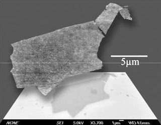
Two overlapping images of the same graphene sheet produced by hydrazine reduction; the top image was produced using atomic force microscopy, while the bottom was produced with scanning electron microscopy. This is the first reported instance of a graphene sheet being large enough for both tests to be run on the same specimen. (Image credit: Vincent Tung, Matthew Allen, Adam Stieg)
Process has already produced the largest graphene sample reported

Graphene is a perfect example of the wonders of nanotechnology, in which common substances are scaled down to an atomic level to uncover new and exciting possibilities.

Graphene is created when graphite — the mother form of all graphitic carbon, which is used to make the pigment that allows pencils to write on paper — is reduced down to a one-atom-thick sheet. Graphene is among the strongest materials known and has an attractive array of benefits.
These sheets — single-layer graphene — have potential as electrodes for solar cells, for use in sensors, as the anode electrode material in lithium batteries and as efficient zero-band-gap semiconductors.

Research on graphene sheets has been restricted, though, due to the difficulty of creating single-layer samples for use in experiments. But in a study published online Nov. 9 in the journal Nature Nanotechnology, researchers from UCLA's California NanoSystems Institute (CNSI) propose a method which can produce graphene sheets in large quantities.

Led by Yang Yang, a professor of materials science and engineering at the UCLA Henry Samueli School of Engineering, and Richard Kaner, a UCLA professor of chemistry and biochemistry, the researchers developed a method of placing graphite oxide paper in a solution of pure hydrazine (a chemical compound of nitrogen and hydrogen), which reduces the graphite oxide paper into single-layer graphene.

Such methods have been studied by others, but this is the first reported instance of using hydrazine as the solvent. The graphene produced from the hydrazine solution is also a more efficient electrical conductor. Field-effect devices display output currents three orders of magnitude higher than previously reported using chemically produced graphene. Kaner and Kang's co-authors on the research were doctoral students Vincent Tung, from Yang's lab, and Matthew Allen, from Kaner's lab.

"We have discovered a route toward solution processing of large-scale graphene sheets," Tung said. "These breakthroughs represent the future of graphene nanoelectronic research."

The coverage of the graphene sheets can be controlled by altering the concentration and composition of the hydrazine solution. This hydrazine method also preserves the integrity of the sheets, producing the largest-area graphene sheet yet reported, 20 micrometers by 40 micrometers. A micrometer is one-millionth of a meter, while a nanometer is one billionth of a meter.

"These graphene sheets are by far the largest produced, and the method allows great control over deposition," Allen said. "Chemically converted graphene can now be studied in depth through a variety of electronic tests and microscopic techniques not previously possible."

"Interdisciplinary research of this sort is a benefit of collaborative institutes like the CNSI," said Kaner, who is also an associate director of the CNSI. "Graphene is a cutting-edge nanomaterial and one which has great potential to revolutionize electronics and many other fields."

There are two methods currently used for graphene production — the drawing method and the reduction method, each with its own drawbacks. In the drawing method, layers are peeled off of graphite crystals until one is produced that is only one-atom thick. When likely graphene suspects are identified from the peeled layers, they must be extensively studied to conclusively prove their identity. In the reduction method, silicon carbide is heated to high temperatures (1100° C) to reduce it to graphene. This process produces a small sample size and is unlikely to be compatible with fabrication techniques for most electronic applications.

"This technology (hydrazine reduction) utilizes a true solution process for graphene, which can dramatically simplify preparing electronic devices," said Yang, who is also faculty director of the Nano Renewable Energy Center at the CNSI. "It thus holds great promise for future large-area, flexible electronics." ###

The California NanoSystems Institute at UCLA is an integrated research center operating jointly at UCLA and the University of California, Santa Barbara, whose mission is to foster interdisciplinary collaborations for discoveries in nanosystems and nanotechnology; train the next generation of scientists, educators and technology leaders; and facilitate partnerships with industry, fueling economic development and promoting the social well-being of California, the United States and the world.

The CNSI was established in 2000 with $100 million from the state of California and an additional $250 million in federal research grants and industry funding. At the institute, scientists in the areas of biology, chemistry, biochemistry, physics, mathematics, computational science and engineering are measuring, modifying and manipulating the building blocks of our world - atoms and molecules.

These scientists benefit from an integrated laboratory culture enabling them to conduct dynamic research at the nanoscale, leading to significant breakthroughs in the areas of health, energy, the environment and information technology. For more information, visit www.cnsi.ucla.edu.

Contact: Mike Rodewald mrodewald@cnsi.ucla.edu 310-267-5883 University of California - Los Angeles

Tuesday, December 23, 2008

New small-scale generator produces alternating current by stretching zinc oxide wires

Prototype Charge Pump

Caption: Image shows a prototype flexible charge pump that generates alternating current as zinc oxide wires are stretched and then released.

Credit: Georgia Tech Photo: Gary Meek. Usage Restrictions: None.
Researchers have developed a new type of small-scale electric power generator able to produce alternating current through the cyclical stretching and releasing of zinc oxide wires encapsulated in a flexible plastic substrate with two ends bonded.

The new "flexible charge pump" generator is the fourth generation of devices designed to produce electrical current by using the piezoelectric properties of zinc oxide structures to harvest mechanical energy from the environment.
Its development was scheduled to be reported November 9, 2008 in the advance online publication of the journal Nature Nanotechnology.

"The flexible charge pump offers yet another option for converting mechanical energy into electrical energy," said Zhong Lin Wang, Regent's professor and director of the Center for Nanostructure Characterization at the Georgia Institute of Technology. "This adds to our family of very small-scale generators able to power devices used in medical sensing, environmental monitoring, defense technology and personal electronics."
The new generator can produce an oscillating output voltage of up to 45 millivolts, converting nearly seven percent of the mechanical energy applied directly to the zinc oxide wires into electricity. The research has been supported by the U.S. Department of Energy, the National Science Foundation, the Air Force Office of Scientific Research and the Emory-Georgia Tech Center for Cancer Nanotechnology Excellence.

Earlier nanowire nanogenerators and microfiber nanogenerators developed by Wang and his research team depended on intermittent contact between vertically-grown zinc oxide nanowires and an electrode, or the mechanical scrubbing of nanowire-covered fibers. These devices were difficult to construct, and the mechanical contact required caused wear that limited how long they could operate. And because zinc oxide is soluble in water, they had to be protected from moisture.
Professor Zhong Lin Wang

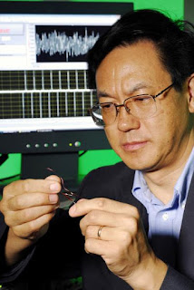
Caption: Georgia Tech Professor Zhong Lin Wang holds a prototype flexible charge pump. The device generates alternating current as zinc oxide wires are stretched and then released.

Credit: Georgia Tech Photo: Gary Meek. Usage Restrictions: None.
"Our new flexible charge pump resolves several key issues with our previous generators," Wang said. "The new design would be more robust, eliminating the problem of moisture infiltration and the wearing of the structures. From a practical standpoint, this would be a major advantage."
Wang with Prototype

Caption: Georgia Tech Professor Zhong Lin Wang holds a prototype flexible charge pump. The device generates alternating current as zinc oxide wires are stretched and then released.

Credit: Georgia Tech Photo: Gary Meek. Usage Restrictions: None.
To boost the current produced, arrays of the flexible charge pumps could be constructed and connected in series. Multiple layers of the generators could also be built up, forming modules that could then be embedded into clothing, flags, building decorations, shoes – or even implanted in the body to power blood pressure or other sensors.

When the modules are mechanically stretched and then released, because of the piezoelectric properties, the zinc oxide material generates a piezoelectric potential that alternately builds up and then is released.
A Schottky barrier controls the alternating flow of electrons, and the piezoelectric potential is the driving force of the charge pump.

"The electrons flow in and out, just like AC current," Wang explained. "The alternating flow of electrons is the power output process."

Constructed with zinc oxide piezoelectric fine wires with diameters of three to five microns and lengths of 200 to 300 microns, the new generator no longer depends on nanometer-scale structures. The larger size was chosen for easier fabrication, but Wang said the principles could be scaled down to the nanometer scale.

"Nanoscale materials are not required for this to work," he said. "Larger fibers work better and are easier to work with to fabricate devices. But the same principle would apply at the nanometer scale."

The wires are grown using a physical vapor deposition method at approximately 600 degrees Celsius. Using an optical microscope, the wires are then bonded onto a polyimide film and silver paste applied at both ends to serve as electrodes. The wires and electrodes were then encased in polyimide to protect them from wear and environmental degradation.

To measure the electric energy generated, the researchers subjected the substrate and attached zinc oxide wires to periodic mechanical bending created by a motor-driven mechanical arm. The bending induced tensile strain which created a piezoelectric potential field along the laterally-packaged wires. That, in turn, drove a flow of electrons into an external circuit, creating the alternating charge and discharge cycle – and corresponding current flow.

Increasing the strain rate increased the magnitude of the output electricity, both in voltage and current. Wang believes the frequency of the current is limited only by the mechanical properties of the polyimide substrate.

The researchers conducted a number of tests to verify that the current measured was produced by the generator – and not an external measurement artifact. Using the same experimental setup, they stretched carbon fibers and Kevlar fibers coated with polycrystalline zinc oxide, and did not observe current flow. The research team also developed two criteria and eight tests for ruling out experimental artifacts, Wang noted.

In addition to Wang, the research team included Rusen Yang and Yong Qin from Georgia Tech and Liming Dai of the Department of Chemical and Materials Engineering at the University of Dayton.

For the future, Wang sees the family of small-scale generators enabling development of a new class self-powered wireless sensing systems. The devices could gather information, store it and transmit the data – all without an external power source.

"Self-powered nanotechnology could be the basis for a new industry," he said. "That's really the only way to build independent systems." ###

Contact: Zhong Lin Wang (404-894-8008); E-mail: (zhong.wang@mse.gatech.edu)

Contact: John Toon jtoon@gatech.edu 404-894-6986 Georgia Institute of Technology Research News

Monday, December 22, 2008

San Diego universities, government and industry rally around local Clean-Tech cluster

Clean Tech Innovation Challenge

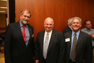
(L-R) Frieder Seible Dean of the UC San Diego Jacobs School of Engineering; San Diego Mayor Jerry Sanders; and Terry Moore, Executive Director, Morrison & Foerster Venture Network recently gave kudos to local researchers making strides in clean technology.
San Diego professors who are developing technologies that will fuel the continued growth of the region's "clean tech cluster" recently received a financial boost through the 2008 Clean Tech Innovation Challenge.

The Clean Tech Innovation Challenge is a partnership between the City of San Diego, UC San Diego's William J. von Liebig Center for Entrepreneurism and San Diego State University (SDSU).
The program is designed to accelerate the commercialization of clean technologies out of university labs as part of the city's goal to promote the growth of the local clean tech industry. Program participants include faculty from UC San Diego, SDSU, University of San Diego and Alliant International University. Qualcomm, Inc. co-sponsored the first grant awards.

"This Clean Tech initiative is an example of how the San Diego community, its universities, local government and the private sector can join forces to create economic growth in the region around technology sectors," said Rosibel Ochoa, the von Liebig Center's acting executive director.

Researchers from UC San Diego and SDSU will receive funding and additional assistance to develop and commercialize new solar technologies, unique ways to convert waste heat to electricity, and novel methods of extracting biodiesel from algae.

"Clean tech is a natural extension of some of the academic and commercial strengths here in San Diego, including electronics, chemistry and biochemistry," said Mike Rondelli, director of the San Diego State Research Foundation.

San Diego Mayor Jerry Sanders and leaders from local universities and San Diego's technology and business communities industry gathered on Oct. 30 to honor the local researchers receiving grants at the 2008 Clean Tech Innovation Challenge Awards Ceremony, which was sponsored by the law firm Morrison & Foerster.

Mayor Jerry Sanders, said during the awards event, "The universities represented here tonight have literally put San Diego on the map. Clean tech is the latest example."

As part of the Clean Tech Innovation Challenge, each researcher nets $50,000, plus business advisory services from the von Liebig Center's consultants. In addition, a team of MBA students from the University of San Diego or Alliant International University will work with each professor in order to conduct market research and create a business plan around the technology. The professors can continue working with their advisor in developing a commercialization plan for the technology and to introduce them to potential funders.

Each grant will support the development of prototypes or the generation of key data that is needed to demonstrate the commercial viability of the technology. The expected timeline for the completion of this program is 12 months. In addition, the awardees have access to other programs like CONNECT's Springboard and the Tech Coast Angeles' Seed track program. They can also seek partnerships with corporations to further develop their technologies.

Ochoa said programs like the von Liebig Center and the Clean Tech Initiative are critical to creating and growing nascent industries. Many researchers, she said, desire to move their inventions into the marketplace but often times lack the resources and funding to make that a reality. This so-called "Valley of Death" is created when federal funding runs out and venture capitalists see the science as too risky to put money into.

"Many of these technologies are so early stage that many investors don't fund them," Ochoa said. "The importance of a program like the Clean Tech Initiative is it allows these researchers to move their technologies further up the value chain so they become attractive to investors or a company to help move them forward.

"UC San Diego is becoming an experimental laboratory for clean technology," she added. "The von Liebig Center is a platform that can be used to demonstrate how these inventions can be turned into commercial technologies."

Ochoa said the Clean Tech Innovation Challenge is unique because it is a private-public partnership.

"The local clean technology industry could be as big as the telecommunications or biotechnology industry, but it requires a concerted effort," she said. "It's important to have this type of public-private partnership to create economic growth and jobs."

Jacques Chirazi, program manager for the City of San Diego Clean Tech Initiative, said the program is right in line with San Diego's famed success of brining innovations from the lab to the marketplace.

"Qualcomm and Cymer are great examples of that," Chirazi said. "We need to continue to tap into the knowledge we have at our local research institutions and universities like UC San Diego and SDSU.

"San Diego has a lot of homegrown technology and science that we can nurture and grow," he added. "The von Liebig Center is a unique model to help this region accomplish that. The center has been recognized as one of the best models in the nation for accelerating research in the nation. The center has a very well designed process of bringing technology from concept to commercialization."

Chirazi said one of the goals of the Clean Tech Innovation Challenge is to inspire innovation in this growing field by encouraging more local researchers, corporations and the San Diego business community to participate in the program.

The following is a brief description of the Clean Tech Innovation Challenge winners and their projects:

Paul Yu, Electrical and Computer Engineering professor, UC San Diego Jacobs School of Engineering
Multiple Quantum Wells for Solar Spectral Concentrator and Optical Energy Transport Technology: In this project, Paul Yu is working on developing new solar-power technologies. In particular, Yu is developing an efficient solar spectral concentrator that will serve as the key component of a technology for transforming a broad-spectral width solar beam into optical energy that can be massively transported via optical fibers to user locations. The solar spectral concentrator can potentially advance the current generation of solar energy collection. Today's photovoltaic (PV) systems are often based upon directly converting solar energy directly into electrical energy. Yu's technology, in contrast, would enable efficient transport and distribution of energy in optical form before final conversion and usage. This will allow for flexible yet direct use of solar energy, and will take advantage of any advances in PV systems. Once established as an alternate way to power up the utilities using solar energy, this proposed technology could be employed broadly world wide.

Yu Qiao, Structural Engineering professor, UC San Diego Jacobs School of Engineering
Developing Ultrahigh-Efficiency Thermal-Energy Harvesting Materials: In this proposed project Qiao and his team are developing unique ways to convert waste heat to electricity by using a nanoporous system. A nanoporous material is a solid that contains nanometer sized pores. This technique re-investigates a mechanism that has long been over-looked and uses it to convert wasted heat to electricity with high efficiency and high power density. The specific goals of this project are to perform comprehensive characterization experiments on nanoporous systems under various conditions; to develop a prototype that can harvest useful electricity from ambient temperature; and to develop a presentation and demonstration set for potential partners, investors, customers, and/or licensees.

John J. Love, Professor, Department of Chemistry and Biochemistry, San Diego State University
Bio-diesel from Cell Membranes: Utilizing Protein Design to Re-Engineer Natural Enzymes for the Extraction of Biodiesel from Cell Membranes: The goal of this project is to process biodiesel from algae. Biodiesel production entails the use of significant amounts of energy for heating, as well as the use of harsh chemicals such as strong bases and/or lye. Love and his team propose to eliminate these costly needs by re-engineering natural protein enzymes such that they efficiently extract fatty acids from membranes and chemically convert them to fatty acid methyl esters (FAMEs), the primary molecules in biodiesel. Examples of membrane sources include bacteria or yeast grown on sugar (glucose) as an energy source or micro-algae grown by way of photosynthesis. ###

Contact: Andrea Siedsma asiedsma@soe.ucsd.edu 858-822-0899 University of California - San Diego

Sunday, December 21, 2008

Gold nanostar shape of the future

Gold Nanostar: Shape of The Future

"To our knowledge, this is the first report of controlled synthesis of gold nanostars with different sizes and shapes and their use as labels for molecular detection. " - Chris Khoury, Graduate student, biomedical engineering

DURHAM, N.C. – Rods, cones, cubes and spheres – move aside. Tiny gold stars, smaller than a billionth of a meter, may hold the promise for new approaches to medical diagnoses or testing for environmental contaminants.

While nanoparticles have been the rage across a wide spectrum of sciences, a new study by Duke University bioengineers indicates that of all the shapes studied to date, stars may shine above all the rest for certain applications.

The key is light, and how that light reflects off the particles. Compared to the other shapes, nanostars can dramatically enhance the reflected light, the Duke scientists found. This increases their potential usefulness as a tracer, label, or contrast agent.

Since the researchers also found that the size and shape of the nanostars affect the spectrum of reflected light, they believe that these tiny nanostars can also be "tuned" to identify particular molecules or chemicals.

"To our knowledge, this is the first report of the development and use of gold nanostars as labels for molecular detection and description of their controlled synthesis with different sizes and shapes" said Chris Khoury, lead author of a paper published on-line in the Journal of Physical Chemistry. Khoury is a graduate student in biomedical engineering working in the laboratory of senior researcher Tuan Vo-Dinh, R. Eugene and Susie E. Goodson Distinguished Professor of Biomedical Engineering and director of The Fitzpatrick Institute for Photonics at Duke.

In the Duke experiments, the nanostars were used in conjunction with a phenomena first described in the 1970s known as surface-enhanced Raman scattering (SERS). When light, usually from a laser, is shined on a sample, the target molecule vibrates and scatters back in its own unique light, often referred to as the Raman scatter. However, this Raman response is extremely weak. When the target molecule is coupled with a metal nanoparticle or nanostructure, the Raman response is greatly enhanced by the SERS effect –often by more than a million times, Vo-Dinh said.

In the early 1980s, while at the Oak Ridge National Laboratory, Tenn., Vo-Dinh and colleagues were among the first to demonstrate that SERS could be put into practical use to detect chemicals including carcinogens, environmental pollutants, and early markers of disease. Now at Duke, Vo-Dinh is pushing the boundaries of the SERS technology by designing a variety of unique types and shapes of metal nanoparticles that can be used as SERS labels for chemical and biomedical detection.

"We are trying to understand which type of nanostructures will give us the optimal signal so we can use them to monitor trace amounts of pollutants or detect diseases in their earliest stage" Vo-Dinh said. "This study is the first demonstration that these nanostars can enhance the effect of SERS to produce strong and unique signatures, like 'optical fingerprints.'"

Khoury "grew" the nanostars by mixing miniscule gold particle seeds in a growth solution. As more gold was added to the solution, protrusions began to sprout from the central core. Additional gold increased the size of the entire particle.

"These experiments demonstrate that it is possible to vary the size and shape of the nanostars in a controlled fashion by adjusting the volume of gold seeds added to the growth solution," Khoury said. "We found that variations in star size changed the reflected light, which hints toward the tuning capabilities that can be exploited by SERS technology."

For such studies, or those involving environmental contaminants, a dye would be attached to the nanostars and mixed with the sample to be tested. The sample would then be placed under a microscope and hit with a burst of laser energy. Sensors would pick up the Raman scattering and interpret the unique optical fingerprint.

Khoury said that nanostars are small enough to pass through cell walls into the interior of the cell, which would make them an effective method for molecular diagnostics. Nanostars could be attached to an antibody to search for antigens, or coupled with a dye to improve the effectiveness of different imaging tests.

While silver enhances the Raman scattering more effectively, gold was chosen as the metallic base of the current nanoparticle because it is a stable metal that doesn't cause immune system reactions within the body. Unlike silver, it also does not oxidize in samples.

Vo-Dinh research group at Duke is currently developing novel techniques for chemical detection and medical diagnostics using SERS. Vo-Dinh said that since each SERS label molecule has its own unique optical fingerprint, theoretically a single probe could be created that could detect an array of different cancers, for example, or different environmental toxins. ###

The research was supported by the National Institutes of Health.

Contact: Richard Merritt richard.merritt@duke.edu 919-660-8414 Duke University

Saturday, December 20, 2008

Stretching silicon: A new method to measure how strain affects semiconductors

Max G. Lagally

Max G. Lagally, Erwin W. Mueller Professor and Bascom Professor of Surface Science.

1109 Engineering Research Building 1500 Engineering Drive Madison, WI 53706 Tel: 608/263-2078 Fax: 608/265-4118 E-mail: lagally@engr.wisc.edu
UW-Madison engineers and physicists have developed a method of measuring how strain affects thin films of silicon that could lay the foundation for faster flexible electronics.

Silicon is the industry standard semiconductor for electronic devices and silicon thin films have the potential to produce faster, more flexible electronics. Researchers have long known that inducing strain into the silicon increases device speed, yet have not fully understood why.

Developed by a team of researchers led by Max Lagally, the Erwin W. Mueller and Bascom Professor of Materials Science and Engineering at UW-Madison, the new method enables the researchers to directly measure the effects of strain on the electronic structure of silicon.
The group published its findings in the Oct. 10 online edition of Physical Review Letters, and the paper will soon appear in the journal's print edition.

Standard strained silicon has so many dislocations and defects that strain measurements are inaccurate, so the research team starts with its own specially fabricated silicon nanomembranes. The team can induce uniform strain in these extremely thin, flexible silicon sheets.

"Imagine if you were to attach a ring and a hook on all four corners and pull equally on all four corners like a trampoline -- it stretches out like that," says Lagally.

As a result, the researchers avoid the defects and variations that make it difficult to study standard strained silicon. Uniform strain allows accurate measurement of its effect on electronic properties.

The researchers drew on the powerful X-ray source at the UW-Madison Synchrotron Radiation Center (SRC), which allowed them to measure conduction bands in strained silicon. To study the energy levels, the researchers needed a wavelength-tunable X-ray source. The SRC also houses a monochromator, a device that enabled the team to choose a precise wavelength, giving their readings the required high energy resolution.

By measuring nanomembranes with different percentages of strain, the researchers have determined the direction and magnitude of shifts in the conduction bands. Their findings have shed light on divergent theories and uncovered some surprising properties. Understanding these properties, and the energy shifts in strained materials, could lead to the improvement of fast, flexible electronic devices.

Capitalizing on its techniques for fabricating silicon nanomembranes, the group hopes to use SRC resources to study strain in other semiconductor materials, as well as to make measurements over smaller areas to study the effects of localized strain.

"The ability to make membranes of various materials, to strain them, and make these measurements will enable us to determine strain-dependent band structure of all kinds of semiconductor materials," says Lagally. ###

Liz Ahlberg, (608) 265-8592, eahlberg@wisc.edu

Contact: Max Lagally lagally@engr.wisc.edu 608-263-2078 University of Wisconsin-Madison

Friday, December 19, 2008

Solar power game-changer: 'Near perfect' absorption of sunlight, from all angles

'Near Perfect' Solar Power

Caption: A new anti-reflective coating developed by researchers at Rensselaer Polytechnic Institute could help to overcome two major hurdles blocking the progress and wider use of solar power. The nanoengineered coating boosts the amount of sunlight captured by solar panels and allows those panels to absorb the entire spectrum of sunlight from any angle, regardless of the sun's position in the sky.

Credit: Rensselaer/Shawn Lin. Usage Restrictions: Include photo credit.
Troy, N.Y. – No matter which way you look at it, the notion of harvesting energy from the sun to power our homes and businesses is more absorbing than ever.

Researchers at Rensselaer Polytechnic Institute have discovered and demonstrated a new method for overcoming two major hurdles facing solar energy. By developing a new antireflective coating that boosts the amount of sunlight captured by solar panels and allows those panels to absorb the entire solar spectrum from nearly any angle, the research team has moved academia and industry closer to realizing high-efficiency, cost-effective solar power.
"To get maximum efficiency when converting solar power into electricity, you want a solar panel that can absorb nearly every single photon of light, regardless of the sun's position in the sky," said Shawn-Yu Lin, professor of physics at Rensselaer and a member of the university's Future Chips Constellation, who led the research project. "Our new antireflective coating makes this possible."

Results of the year-long project are explained in the paper "Realization of a Near Perfect Antireflection Coating for Silicon Solar Energy," published this week by the journal Optics Letters.

An untreated silicon solar cell only absorbs 67.4 percent of sunlight shone upon it — meaning that nearly one-third of that sunlight is reflected away and thus unharvestable. From an economic and efficiency perspective, this unharvested light is wasted potential and a major barrier hampering the proliferation and widespread adoption of solar power.

After a silicon surface was treated with Lin's new nanoengineered reflective coating, however, the material absorbed 96.21 percent of sunlight shone upon it — meaning that only 3.79 percent of the sunlight was reflected and unharvested. This huge gain in absorption was consistent across the entire spectrum of sunlight, from UV to visible light and infrared, and moves solar power a significant step forward toward economic viability.

Lin's new coating also successfully tackles the tricky challenge of angles.

Most surfaces and coatings are designed to absorb light — i.e., be antireflective — and transmit light — i.e., allow the light to pass through it — from a specific range of angles. Eyeglass lenses, for example, will absorb and transmit quite a bit of light from a light source directly in front of them, but those same lenses would absorb and transmit considerably less light if the light source were off to the side or on the wearer's periphery.

This same is true of conventional solar panels, which is why some industrial solar arrays are mechanized to slowly move throughout the day so their panels are perfectly aligned with the sun's position in the sky. Without this automated movement, the panels would not be optimally positioned and would therefore absorb less sunlight. The tradeoff for this increased efficiency, however, is the energy needed to power the automation system, the cost of upkeeping this system, and the possibility of errors or misalignment.

Lin's discovery could antiquate these automated solar arrays, as his antireflective coating absorbs sunlight evenly and equally from all angles. This means that a stationary solar panel treated with the coating would absorb 96.21 percent of sunlight no matter the position of the sun in the sky. So along with significantly better absorption of sunlight, Lin's discovery could also enable a new generation of stationary, more cost-efficient solar arrays.

"At the beginning of the project, we asked 'would it be possible to create a single antireflective structure that can work from all angles?' Then we attacked the problem from a fundamental perspective, tested and fine-tuned our theory, and created a working device," Lin said. Rensselaer physics graduate student Mei-Ling Kuo played a key role in the investigations.

Typical antireflective coatings are engineered to transmit light of one particular wavelength. Lin's new coating stacks seven of these layers, one on top of the other, in such a way that each layer enhances the antireflective properties of the layer below it. These additional layers also help to "bend" the flow of sunlight to an angle that augments the coating's antireflective properties. This means that each layer not only transmits sunlight, it also helps to capture any light that may have otherwise been reflected off of the layers below it.

The seven layers, each with a height of 50 nanometers to 100 nanometers, are made up of silicon dioxide and titanium dioxide nanorods positioned at an oblique angle — each layer looks and functions similar to a dense forest where sunlight is "captured" between the trees. The nanorods were attached to a silicon substrate via chemical vapor disposition, and Lin said the new coating can be affixed to nearly any photovoltaic materials for use in solar cells, including III-V multi-junction and cadmium telluride. ###

Along with Lin and Kuo, co-authors of the paper include E. Fred Schubert, Wellfleet Senior Constellation Professor of Future Chips at Rensselaer; Research Assistant Professor Jong Kyu Kim; Research Associate Yong Sung Kim; physics graduate student David Poxson; and electrical engineering graduate student Frank Mont.

Funding for the project was provided by the U.S. Department of Energy's Office of Basic Energy Sciences, as well as the U.S. Air Force Office of Scientific Research.

Contact: Michael Mullaney mullam@rpi.edu 518-276-6161 Rensselaer Polytechnic Institute

Thursday, December 18, 2008

New nanocluster to boost thin films for semiconductors

Graduate student Zachary Mensinger, left, talks with co-authors Lev N. Zakharov, center, and Darren Johnson

Caption: Graduate student Zachary Mensinger, left, talks with co-authors Lev N. Zakharov, center, and Darren Johnson in Zakharov's lab in the underground Lorry I. Lokey Laboratories at the University of Oregon.

Credit: Photo by Jim Barlow. Usage Restrictions: None.
University of Oregon, Oregon State discovery speeds production and yields; may lead to greener process.

Oregon researchers have synthesized an elusive metal-hydroxide compound in sufficient and rapidly produced yields, potentially paving the way for improved precursor inks that could boost semiconductor capabilities for large-area applications.
The key to a "bottom-up" production of possibly the first heterometallic gallium-indium hydroxide nanocluster was the substitution of nitroso-butylamine as an additive in place of nitrosobenzene.

The substitute was identified during a comprehensive screening of potential alternatives by Zachary L. Mensinger, a doctoral student in the lab of University of Oregon chemist Darren W. Johnson. The additive acts to optimize and speed crystallization, allowing for reaction yields up to 95 percent. Comparable compounds traditionally made under caustic conditions often take months or even years to crystallize and result in low yields.

"The benefit is that we can predictably control the ratio of gallium and indium in these structures at molecular levels, which can result in the same control in the fabrication of semiconductor thin films," Johnson said. "We can tailor the properties for specific applications or for different performance levels."

Six University of Oregon and Oregon State University collaborators, working under the umbrella of the Oregon Nanoscience and Microtechnologies Institute (ONAMI), a state signature research center, describe their findings a paper to appear in the German Chemical Society's journal Angewandte Chemie (Applied Chemistry) International. The research, published early online, also was performed under the auspices of a new National Science Foundation-funded Center for Green Materials Chemistry, operated jointly by the two Oregon universities.

"Researchers working in the solid-state materials community are looking at these kinds of nanoclusters as precursors for thin films and other advanced materials, but you typically cannot get them in high enough yields," said Johnson, who also is a member of the UO's Materials Science Institute. "Our synthesis, however, allows for gram-scale quantities."

The results represent a significant breakthrough in the way liquids are produced for semiconductor fabrication, said co-author Douglas A. Keszler, distinguished professor of chemistry at Oregon State and adjunct UO chemistry professor. "We now have new methods for pushing printed inorganic electronics to higher levels of performance within a useful class of materials."

Researchers in Johnson's lab have been experimenting with low-temperature production of a series of such heterometallic nanoclusters, which consist of 13 atoms and contain two different metals in the metal 13 framework, which may prove desirable for long-term applications in solid-state electronics. The nanocluster identified in the paper is labeled a Ga7In6 hydroxide.

"We're starting from a bottom-up approach, in that we can make these with the ratios we desire already built in," Mensinger said. "Using this nitroso compound, we get a higher yield and at a larger scale. I screened several of these compounds to narrow down the best choice. We can also re-use the nitroso compound. It is still present at the end of the reaction, so we can remove it and use it in future reactions."

While the nitroso compound produces usable amounts of nanoclusters for potential semiconductor applications and is reusable in subsequent production, it is toxic, Johnson noted. "It is great because it allowed us to make these clusters that had never been made before, but it is not truly a green-chemistry method," Johnson said. "We're looking at how it works and hope to replace it with a more benign reagent." ###

Co-authors with Mensinger, Johnson and Keszler on the paper were Jason T. Gatlin and Lev N. Zakharov, both of the University of Oregon, and Stephen T. Meyers, a graduate student of Keszler's at OSU.

The National Science Foundation helped support the research through a CAREER award to Johnson and an Integrative Graduate Education and Research Training grant. Additional funding came from the Research Corp. through a 2006 Cottrell Scholar award to Johnson and Army Research Laboratory funds provided through ONAMI.

About the University of Oregon

The University of Oregon is a world-class teaching and research institution and Oregon's flagship public university. The UO is a member of the Association of American Universities (AAU), an organization made up of 62 of the leading public and private research institutions in the United States and Canada. Membership in the AAU is by invitation only. The University of Oregon is one of only two AAU members in the Pacific Northwest.

Sources: Darren Johnson, assistant professor of chemistry, 541-346-1695, dwj@uoregon.edu; Douglas Keszler, distinguished professor of chemistry, Oregon State University, 541-737-6736, Douglas.Keszler@oregonstate.edu

Links: Contact: Jim Barlow jebarlow@uoregon.edu 541-346-3481 University of Oregon

Wednesday, December 17, 2008

Ultrafast lasers give CU-Boulder researchers a snapshot of electrons in action

laser beam excites dinitrogen tetraoxide molecules, or N2O4

A laser beam first excites dinitrogen tetraoxide molecules, or N2O4, inducing large vibrations. A second laser beam then generates X-rays from the vibrating molecules.
In the quest to slow down and ultimately understand chemistry at the level of atoms and electrons, University of Colorado at Boulder and Canadian scientists have found a new way to peer into a molecule that allows them to see how its electrons rearrange as the molecule changes shape.

Understanding how electrons rearrange during chemical reactions could lead to breakthroughs in materials research and in fields like catalysis and alternative energy, according to CU-Boulder physics professors and JILA fellows Margaret Murnane and Henry Kapteyn,
who led the research efforts with scientist Albert Stolow of the Canadian National Research Council's Steacie Institute for Molecular Sciences.

"The Holy Grail in molecular sciences would be to be able to look at all aspects of a chemical reaction and to see how atoms are moving and how electrons are rearranging themselves as this happens," Murnane said. "We're not there yet, but this is a big step toward that goal."

To be able to chart a chemical reaction, scientists need to be able to see how bonds are formed or broken between atoms in a molecule during chemical reactions. But only extremely limited tools are available to view the rapidly changing electron cloud that surrounds a molecule as the atoms move around, Murnane said. Changes in the electron cloud can happen on timescales of less than a femtosecond, or one quadrillionth of a second, representing some of the fastest processes in the natural world.

In a paper to appear in the Oct. 30 issue of Science Express, the online version of the journal Science, the CU team describes how they shot a molecule of dinitrogen tetraoxide, or N2O4, with a short burst of laser light to induce very large oscillations within the molecule. They then used a second laser to produce an X-ray, which was used to map the electron energy levels of the molecule, and most importantly, to understand how these electron energy levels rearrange as the molecule changes its shape, according to Kapteyn.

"This is a fundamentally new way of looking at molecules," Kapteyn said. "This process allowed us to freeze the motion of electrons in a system, and to capture their dizzying dance."

The researchers describe their process of stretching the N2O4 molecule as being similar to pulling on a Slinky toy and then letting it go and watching it vibrate. They used the N2O4 molecule because it vibrates more slowly compared to other molecules, allowing them to observe the physical processes under way.

In many ways, molecules are like tiny masses connected by tiny springs of differing strengths, Murnane said. These springs are the chemical bonds, made up of shared electrons, which hold all matter together. In this experiment they used ultrafast laser pulses to "twang" these springs, making the nanoscale molecular Slinkies vibrate. However, unlike real springs, when researchers vibrate the molecules their properties can change, she said.

Being able to watch and understand why the electrons did what they did is very useful in fields like alternative energy, according to the researchers.

"If we understand the nature of these processes, in the future we can then translate that knowledge into better technology, such as creating more efficient light-harvesting molecules or catalysis or perhaps even solar cells," Stolow said. ###

The research was completed by an international team with JILA Research Associate Wen Li as the paper's corresponding author. He worked with CU-Boulder physics graduate students Xibin Zhou and Robynne Lock as well as Serguei Patchkovskii and Stolow of the Steacie Institute for Molecular Sciences in Ottawa, Canada.

JILA is a joint institute of CU-Boulder and the National Institute of Standards and Technology.

Contact: Margaret Murnane margaret.murnane@colorado.edu 303-492-7839 University of Colorado at Boulder. Henry Kapteyn, 303-492-8198, Greg Swenson, 303-492-3113.

Tuesday, December 16, 2008

Nanoscale dimensioning is fast, cheap with new NIST optical technique

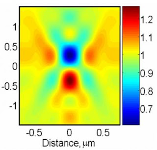
This schematic shows how a TSOM image is acquired.

Caption: This schematic shows how a TSOM image is acquired. Using an optical microscope, several images of a 60 nanometer gold particle sample (shown in red) are taken at different focal positions and stacked together.

Credit: NIST. Usage Restrictions: None.
A novel technique* under development at the National Institute of Standards and Technology (NIST) uses a relatively inexpensive optical microscope to quickly and cheaply analyze nanoscale dimensions with nanoscale measurement sensitivity. Termed “Through-focus Scanning Optical Microscope” (TSOM) imaging, the technique has potential applications in nanomanufacturing, semiconductor process control and biotechnology.

Optical microscopes are not widely considered for checking nanoscale (below 100 nanometers) dimensions because of the limitation imposed by wavelength of light—you can’t get a precise image with a probe three times the object’s size. NIST researcher Ravikiran Attota gets around this, paradoxically, by considering lots of “bad” (out-of-focus) images.
“This imaging uses a set of blurry, out-of-focus optical images for nanometer dimensional measurement sensitivity,” he says. Instead of repeatedly focusing on a sample to acquire one best image, the new technique captures a series of images with an optical microscope at different focal positions and stacks them one on top of the other to create the TSOM image. A computer program Attota developed analyzes the image.

While Attota believes this simple technique can be used in a variety of applications, he has worked with two. The TSOM image can compare two nanoscale objects such as silicon lines on an integrated circuit. The software “subtracts” one image from the other. This enables sensitivity to dimensional differences at the nanoscale—line height, width or side-wall angle. Each type of difference generates a distinct signal.
TSOM has also been theoretically evaluated in another quality control application. Medical researchers are studying the use of gold nanoparticles to deliver advanced pharmaceuticals to specific locations within the human body. Perfect size will be critical. To address this application, a TSOM image of a gold nanoparticle can be taken and compared to a library of simulated images to obtain “best-match” images with the intent of determining if each nanoparticle passes or fails.

This new imaging technology requires a research-quality optical microscope, a camera and a microscope stage that can move at preset distances.
several images of a 60 nanometer gold particle sample

Caption: Using an optical microscope, several images of a 60 nanometer gold particle sample are taken at different focal positions and stacked together. This computer-created image shows the resultant TSOM image.

Credit: NIST. Usage Restrictions: None.
“The setup is easily under $50,000, which is much less expensive than electron or probe microscopes currently used for measuring materials at the nanoscale,” Attota explains. “This method is another approach to extend the range of optical microscopy from microscale to nanoscale dimensional analysis.” So far, sensitivity to a 3 nm difference in line widths has been demonstrated in the laboratory. ###

* R. Attota, T.A. Germer and R.M. Silver. Through-focus scanning-optical-microscope imaging method for nanoscale dimensional analysis, Optics Letters 33, 1990 (2008).

Contact: Evelyn Brown evelyn.brown@nist.gov 301-975-5661 National Institute of Standards and Technology (NIST)

Monday, December 15, 2008

Sniffing out a better chemical sensor

Sniffing Out a Better Chemical Sensor

Caption: NIST researchers have developed a new approach for 'electronic noses.' Comprised of 16 microheater elements and eight types of sensors, the tiny device could be a potent tool for applications such as sniffing out nerve agents, environmental contaminants, and trace indicators of disease, in addition to monitoring industrial processes and aiding in space exploration.

Credit: NIST. Usage Restrictions: None.
Marrying a sensitive detector technology capable of distinguishing hundreds of different chemical compounds with a pattern-recognition module that mimics the way animals recognize odors, researchers at the National Institute of Standards and Technology (NIST) have created a new approach for “electronic noses.”
Described in a recent paper,* their electronic nose is more adept than conventional methodologies at recognizing molecular features even for chemicals it has not been trained to detect and is also robust enough to deal with changes in sensor response that come with wear and tear. The detector could be a potent tool for applications such as sniffing out nerve agents, environmental contaminants, and trace indicators of disease, in addition to monitoring industrial processes and aiding in space exploration.

In animals, odorant molecules in the air enter the nostrils and bind with sensory neurons in the nose that convert the chemical interactions into an electrical signal that the brain interprets as a smell. In humans, there are about 350 types of sensory neurons and many copies of each type; dogs and mice have several hundreds more types of sensory neurons than that. Odor recognition proceeds in a step-by-step fashion where the chemical identity is gradually resolved: initial coarse information (e.g. ice-cream is fruit-flavored vs. chocolate) is refined over time to allow finer discrimination (strawberry vs. raspberry). This biological approach inspired the researchers to develop a parallel “divide and conquer” method for use with the electronic nose.

The technology is based on interactions between chemical species and semiconducting sensing materials placed on top of MEMS microheater platforms developed at NIST. (See “NIST ‘Microhotplate’ May Help Search for Extraterrestrial Life,” NIST Tech Beat, Oct., 2001.) The electronic nose employed in the current work is comprised of eight types of sensors in the form of oxide films deposited on the surfaces of 16 microheaters, with two copies of each material. Precise control of the individual heating elements allows the scientists to treat each of them as a collection of “virtual” sensors at 350 temperature increments between 150 to 500 °C, increasing the number of sensors to about 5,600. The combination of the sensing films and the ability to vary the temperature gives the device the analytical equivalent of a snoot full of sensory neurons.

Much like people detect and remember many different smells and use that knowledge to generalize about smells they haven’t encountered before, the electronic nose also needs to be trained to recognize the chemical signatures of different smells before it can deal with unknowns. The great advantage of this system, according to NIST researchers Barani Raman and Steve Semancik, is that you don’t need to expose the array to every chemical it could come in contact with in order to recognize and/or classify them. Breaking the identification process down into simple, small, discrete steps using the most information rich data also avoids ‘noisy’ portions of the sensor response, thereby incorporating robustness against the effects of sensor drift or aging.

The researchers say that they are continuing to work on applications involving rapid identification of chemicals in unknown backgrounds or in a complex cocktail. ###

* B. Raman, J. L. Hertz, K. D. Benkstein and S. Semancik. Bioinspired methodology for artificial olfaction. Analytical Chemistry. Published online Oct. 15, 2008.

Contact: Mark Esser mark.esser@nist.gov 301-975-8735 National Institute of Standards and Technology (NIST)

Tags: or and