Wednesday, January 31, 2007

Health and safety impacts of nanotechnology

Researchers probe health and safety impacts of nanotechnology

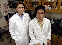
GAINESVILLE, Fla. — University of Florida engineering student Maria Palazuelos is working on nanotechnology, but she’s not seeking a better sunscreen, tougher golf club or other product — the focus of many engineers in the field.

Instead, Palazuelos, a doctoral student in chemical engineering, is probing the potentially harmful effects of nanotechnology by testing how ultra-small particles may adversely affect living cells, organisms and the environment. But this is no scene from Michael Crichton’s novel “Prey” about nanotechnology run amok. Rather, this is a real-world endeavor grounded in solid science.

“We don’t want to look back in 50 years if something bad has happened and say, ‘why didn’t we ask these questions?’” Palazuelos said.

Palazuelos is a member of a small interdisciplinary group of UF faculty members and students, the UF Nanotoxicology Group, whose work is rapidly becoming more timely as manufacturers increasingly turn to the super-small tubes, cylinders and other nanoparticles at the heart of nanotechnology.

There are already more than 400 companies worldwide that tap nanoparticles and other forms of nanotechnology, and regulatory agencies such as the Environmental Protection Agency, the Food and Drug Administration and the Occupational Health and Safety Administration are closely examining whether new regulations are needed to guard against potentially harmful but currently unknown effects, said Kevin Powers, associate director of UF’s National Science Foundation and Particle Engineering Research Center. These agencies are turning to university researchers for help in making those kinds of determinations, he said.

“Before we start producing these materials in large quantities to go into everyday products, we should know what effect they have on our health and the environment,” he said.

The UF group consists of about 10 faculty members and a half-dozen students from UF’s engineering, medical and veterinary colleges. With funding from UF and agencies including the EPA, National Science Foundation and the U.S. Air Force, the researchers have at least eight projects aimed at answering questions ranging from how nanoparticles affect fish to whether nanoparticles can penetrate skin. The researchers have presented several lectures at conferences and have several papers published or undergoing review.

Powers said the health and environmental effects of common metals and materials are well-known. The question for the researchers is whether the effects change when the metals and materials take the form of nanoparticles – and whether these nanoparticles become more or less hazardous based on shape and size. “It’s complicated,” he said. “In many cases, we lack basic knowledge of the properties and the behavior of the particles themselves.”

Palazuelos is investigating what happens to living cells when confronted with aluminum nanoparticles. For the type of cells she has tested, the cells can readily absorb the aluminum nanoparticles, and there is a correlation between size, shape and toxicity. That said, Palazuelos stressed that it is far too early to conclude that aluminum nanoparticles are harmful to human health. Epidemiological studies evaluating years of exposure to aluminum in foundry workers and welders have not shown dramatic health effects as long as basic safety and exposure guidelines are followed, she said.

“It is a long way from isolated tissue studies to the extrapolation of these results to human health,” she said. “However, a fundamental understanding of the nanoparticle-cell interactions will be very useful in this field.”

Copper and some other metals are known to be toxic to fish and other aquatic wildlife. UF toxicologist David Barber is investigating whether nanoparticles made of these metals are more toxic than standard soluble forms of the metals. As part of his research, he has exposed zebra fish, a species commonly used in laboratory tests, to various concentrations of copper nanoparticles and compared the results with those induced by copper sulfate.

His results so far show that the 30-nanometer, spherical copper nanoparticles are lethal to zebra fish, though less toxic than copper sulfate. However, the way the copper nanoparticles cause damage is different. “Both of them are causing lethality by affecting the gill,” Barber said. “The lesion is slightly different, and the gene expression response in the gills is very different.”

Barber said he hopes to determine whether the size or shape of the nanoparticle is key to its effects. If that’s the case, it could mean a lot of work ahead for regulatory agencies.

“Typically, when you test a chemical, the response is the same regardless of formulation. Aspirin is always aspirin,” Barber said. “If all of a sudden every time you change the size or the shape of a nanoparticle you have to retest it; that’s a lot of testing.”

Credits Writer Aaron Hoover, ahoover@ufl.edu, 352-392-0186, Source Kevin Powers, kpowers@erc.ufl.edu, 352-846-1194, Source David Barber, dbarber@ufl.edu, 352-392-4700, ext. 5540, © University of Florida, Gainesville, FL 32611; (352) 392-3261.

Technorati Tags: or and or and or and or and or and or and or and or

Nano-Hazard Symbol Contest

Winners of Nano-Hazard Symbol Contest Announced at World Social Forum, Nairobi, Kenya

Nano-Hazard Symbol
An estimated 30,000 people gathered at the World Social Forum in Nairobi this week where participants had a chance to vote for their favorite Nano-Hazard Symbol – a design that warns of the presence of engineered nanomaterials (1 nanometer = 1 billionth of a meter).

The winners of the international graphic design competition were announced today. The winning designs were submitted by: Dimitris Deligiannis (Greece), Shirley Gibson (Scotland), and Kypros Kyprianou (England).

“Tiny tech is no small matter – there was intense competition to design a nano-hazard symbol, and enormous interest in Nairobi,” said Pat Mooney of ETC Group. “We ended up with three winners who were virtually tied for first place,” explained Mooney.

The competition netted 482 unique designs from 24 countries. An independent panel of judges selected 16 finalists that appeared on the ballot in Nairobi. (The 16 finalists can be found here)

The winning designs will be submitted to international standard-setting bodies responsible for hazard characterisation and could be used as a label on product-packaging or workroom walls. Because of their extremely small size and large surface area, nano-scale particles may be more reactive and more toxic than larger particles of the same substance. Even though hundreds of products containing engineered nanoparticles are on the market, the toxicology of nanoparticles is largely unknown.

(More information on the competition, along with the list of judges, can be found here: etcgroup.org/nanohazard)

ETC Group, News Release, Wednesday, 24 January 2007 etcgroup.org

All 482 design submissions can be viewed here. and The 16 finalists for the Nairobi-phase of the contest here.

ETC contact information: Pat Mooney and Kathy Jo Wetter of ETC Group are attending the World Social Forum. We have a booth at the WSF venue and / or can be reached by email: Pat Mooney: etc@etcgroup.org , Kathy Jo Wetter: kjo@etcgroup.org

Hope Shand (US) hope@etcgroup.org tel: 919 960-5767
Silvia Ribeiro (Mexico) silvia@etcgroup.org tel: +52 5555 6326 64

Technorati Tags: or and or and or and or and or and or and or and or

Tuesday, January 30, 2007

DNA sequencing technologies

Faster, low cost sequencing technologies needed to drive era of personalized medicine High Resolution Image, 300 DPI, PODCAST FOR THIS ARTICLE

Description: Image of DNA Double Helix, Courtesy: National Human Genome Research Institute.DNA testing is transforming health care and medicine, but current technologies only give a snapshot of an individual's genetic makeup. Any patient wanting a complete picture of their inherited DNA, or genome,
would drop their jaw at the sight of the bill -- to the current tune of $10 million or more charged for every human or mammalian-sized genome sequenced.

Now, with a grant award from the National Human Genome Research Institute (NHGRI), scientists at the Biodesign Institute at Arizona State University are expanding efforts to dramatically lower the cost of DNA sequencing.
The NHGRI, part of the National Institutes of Health (NIH), has set an ambitious target of $1,000 or less - a cost 10,000 times lower than current technology - to make genome sequencing a routine diagnostic tool in medical care. The reduced cost may allow doctors to tailor medical treatments to an individual's genetic profile for diagnosing, treating, and ultimately preventing many common diseases such as cancer, heart disease, diabetes and obesity.
ASU chemist Peiming Zhang and his collaborator Jian Gu have been awarded a $897,000 grant under this program for an ambitious DNA sequencing project that combines physics, chemistry and nanotechnology with engineering. The researchers have been charged with the daunting task of shrinking down the 13 year, $2.7 billion Human Genome Project to days.

"If you want to develop a technology to sequence an individual genome for $1,000, you have to think about using nanotechnology," said Zhang, associate research professor in the Center for Single Molecule Biophysics at the Biodesign Institute. "The technology is available now to pioneer a new approach to sequencing."

Much like the computer industry, DNA sequencing technology is driven by the mantra of faster, cheaper and more reliable. In the past generation, sequencing costs have fallen 100-fold, from roughly a dollar a DNA base to a penny, but are still far out of reach for the public.

Zhang's technological vision would enable scientists to sequence billions of base pairs of DNA in a single day. This is the size of an average mammalian genome and is approximately 10,000 times more bases per day than can be sequenced using current technologies. By increasing the speed of sequencing and reducing its cost, genetic research may develop a more significant role in everyday medical practice.

In Zhang's sequencing project, billions of base pairs of genomic DNA could be sequenced on a single, cookie crumb-sized one centimeter by one centimeter chip. The technique uses hybridization, a process of joining two complementary strands of DNA, to sequence DNA by applying a sample to single stranded DNA probes attached to a chip.

An atomic force microscope (AFM), like a caffeinated speed reader, can then rapidly scan the surface of the chip to see where DNA from the sample has hybridized to the probes. Wherever sample DNA binds to the probes, the sequence is registered.

"Traditional approaches to sequencing by hybridization are limited by the number of probes that can be placed on a chip," said Jian Gu, a research staff member in the Center for Applied NanoBioscience at the Biodesign Institute and co-leader of the project.

By using nanoprinting techniques developed by Gu, the researchers hope to increase the number probes they can fit on a chip. "Right now, we have a mechanical printing technology that could put down billions of probes on a chip surface at very low cost," said Gu.

It is estimated that a single base pair can be sequenced for every DNA probe, which means that optimizing the nanoprinting process is critical to the goal of a $1,000 genome, according to Zhang.

The researchers' first goal is a proof of principal for their approach. They plan to synthesize a universal DNA nanoarray on a 100 micrometer by 100 micrometer chip, about the size of a dust mite, by 2009.

The award to Zhang and his team was one of nine grants given by the NIH to achieve the $1,000 genome goal. Zhang's effort also joins two other ASU research teams, led by Stuart Lindsay and Peter Williams, who have more than $2 million in other DNA sequencing projects funded at ASU.

"There are currently only 36 grants in the entire NHGRI sequencing program, so it's quite remarkable that ASU has three of them, which is almost 10 percent of the program," Williams said.

Williams, professor of chemistry and biochemistry, is working on a $100,000 genome project, part of the five-year goal of the NHGRI to drop the current price to a hundredth of the cost. His goal is to selectively sequence genes known to be involved in disease in a matter of hours, and for a few hundred dollars.

Lindsay, who is director of the Biodesign Institute's Center for Single Molecule Biophysics, is engaged in a different separately funded $1,000 genome project. Lindsay is threading DNA through a molecular ring, in this case a sugar called cyclodextrin, that can read the DNA sequence by measuring the differences in friction as the molecule is pulled through the ring. ###

The Biodesign Institute at ASU combines research in biology, engineering, information technology, and cognitive science to accelerate discoveries into beneficial uses. The institute currently is pursuing innovations in healthcare, national security and environmental sustainability. For information, visit www.biodesign.asu.edu or call (480) 727-8322.

Contact: Joe Caspermeyer joseph.caspermeyer@asu.edu 480-727-0369 Arizona State University

Technorati Tags: or and or and or and or and or and or and or and or

Monday, January 29, 2007

Coated nanoparticles drug-delivery

Coated nanoparticles solve sticky drug-delivery problem, Researchers take cues from viruses to get treatment through body's protective. PODCAST FOR THIS ARTICLE.

Caption: In a Johns Hopkins chemical and biomolecular engineering lab, Justin Hanes, an associate professor, and doctoral student Samuel K. Lai tested coated nanoparticles that could deliver medications through the body's sticky mucus layers. Credit: Will Kirk/JHU, Usage Restrictions: None.The layers of mucus that protect sensitive tissue throughout the body have an undesirable side effect: they can also keep helpful medications away. To overcome this hurdle,
Johns Hopkins researchers have found a way to coat nanoparticles with a chemical that helps them slip through this sticky barrier.

During experiments with these coated particles, the researchers also discovered that mucus layers have much larger pores than previously thought, providing a doorway that should allow larger and longer-acting doses of medicine to reach the protected tissue.

The team's findings were reported this week in the Early Online Edition of Proceedings of the National Academy of Sciences.
The discoveries are important because mucus layers, which trap and help remove pathogens and other foreign materials, can block the localized delivery of drugs to many parts of the body, including the lungs, eyes, digestive tract and female reproductive system. Because of these barriers, doctors often must prescribe pills or injections that send drugs through the entire body,Caption: Using high-resolution video microscopy and computer software, doctoral student Samuel K. Lai and Justin Hanes, associate professor of chemical and biomolecular engineering at Johns Hopkins, were able to track their coated nanoparticles as the potential medication carriers made their way through a mucus layer. Credit: Will Kirk/JHU, Usage Restrictions: None.
an approach that can lead to unwanted side effects or doses that are too weak to provide effective treatment.

"Mucus barriers evolved to serve a helpful purpose: to keep things out," said Justin Hanes, an associate professor of chemical and biomolecular engineering who supervised the research. "But if you want to deliver medicine in a microscopic particle, they can also keep the drugs from getting through. We've found a way to keep helpful nanoparticles from sticking to mucus, and we learned that the openings in the mucus 'mesh' are much larger than most people expected. These findings set the stage for a new generation of nanomedicines that can be delivered directly to the affected areas."

To get its particles past the mucus, Hanes' team studied an unlikely model: viruses. Earlier research led by Richard Cone, a professor in the Department of Biophysics at Johns Hopkins, had established that some viruses are able to make their way through the human mucus barrier. Hanes and his colleagues decided to look for a chemical coating that might mimic the characteristics of a virus.

"We found that the viruses that got through had surfaces that were attracted to water, and they had a net neutral electrical charge," said Samuel K. Lai, a Johns Hopkins chemical and biomolecular engineering doctoral student from Canada and Hong Kong who was lead author of the journal article. "We thought that if we could coat a drug-delivery nanoparticle with a chemical that had these characteristics, it might not get stuck in the mucus barrier."

To make their nanoparticles behave like viruses, the researchers coated them with polyethylene glycol, PEG, a non-toxic material commonly used in pharmaceuticals. PEG dissolves in water and is excreted harmlessly by the kidneys.

The researchers also considered the size of their nanoparticles. Previous studies indicated that even if nanoparticles did not stick to the mucus, they might have to be smaller than 55 nanometers wide to pass through the tiny openings in the human mucus mesh. (A human hair is roughly 80,000 nanometers wide.) Using high-resolution video microscopy and computer software, the researchers discovered that their PEG-coated 200-nanometer particles could slip through a barrier of human mucus.

They then conducted further tests to see how large their microscopic drug carriers could be before they got trapped in the mesh. Larger nanoparticles are more desirable because they can release greater amounts of medicine over a longer period of time. "We wanted to make the particles as large as possible," said Hanes, who also serves as director of therapeutics for the Institute for NanoBioTechnology at Johns Hopkins. "The shocking thing was how fast the particles that were 500 nanometers wide moved through the mucus mesh. The work suggests that the openings in the mucus barrier are much larger than originally expected by most. And we were also surprised to find that the larger nanoparticles (200 and 500 nanometers wide) actually moved through the mucus layer more quickly than the smaller ones (100 nanometers wide)."

This has important implications, Hanes said, because a 500-nanometer particle can be used to deliver medicine to a targeted area, released over periods of days to weeks. Larger particles also allow a wider array of drug molecules to be efficiently encapsulated. He and his colleagues believe this system has great potential in the delivery of chemotherapy, antibiotics, nucleic acids and other treatment directly to the lungs, gastrointestinal tract and cervicovaginal tract. ###

Through Johns Hopkins Technology Transfer, the team has applied for patents covering this process.

In addition to Lai, Hanes and Cone, co-authors of the PNAS paper included D. Elizabeth O'Hanlon and Suzanne Harrold, doctoral students in the Johns Hopkins Department of Biophysics in the Krieger School of Arts and Sciences; Stan T. Man, a former visiting research scientist in the Johns Hopkins Department of Chemical and Biomolecular Engineering in the Whiting School of Engineering; and Ying-Ying Wang, who contributed to the research as a Johns Hopkins undergraduate and who is now a graduate student in the university's Department of Biomedical Engineering.

Lai's participation was partially supported by a scholarship from the Natural Science and Engineering Research Council of Canada.

Digital photos of the researchers available; contact Phil Sneiderman.

Contact: Phil Sneiderman prs@jhu.edu 443-287-9960 Johns Hopkins University

Related Links: Justin Hanes' Lab Page: jhu.edu/~cheme/hanes/ Department of Chemical and Biomolecular Engineering: jhu.edu/chbe/ Richard Cone's Lab Page: biophysics.jhu.edu/cone/ Institute for NanoBioTechnology ay Johns Hopkins: inbt.jhu.edu/

Technorati Tags: or and or and or and or and or and or and or and or

Sunday, January 28, 2007

'Biomimetic Technologies' soft-bodied robots

'Biomimetic Technologies' project will create first soft-bodied robots, New devices could be used in operating rooms, space stations. KINEMATIC STUDIES MPG of Image. PODCAST FOR THIS ARTICLE.

MEDFORD/SOMERVILLE, Mass. -- While robots have moved from the realm of science fiction to a myriad of real-life uses, the potential of the "hard-bodied" robots of the 21st century
remains limited by their stiff construction and lack of flexibility. A group of researchers at Tufts University has launched a multidisciplinary initiative focused on the science and engineering of a new class of robots that are completely soft-bodied. These devices will make possible advances in such far flung arenas as medicine and space exploration.

Barry Trimmer, professor of biology, and David Kaplan, professor of biomedical engineering, are co-directors of the Biomimetic Technologies for Soft-bodied Robots project, which represents a consortium of seven Tufts faculty members from five departments in the School of Engineering and the School of Arts and Sciences. The project has just been awarded a grant of $730,000 from the W.M. Keck Foundation.

According to Kaplan, the project will bring together biology, bioengineering and micro/nano fabrication. "Our overall goal is to develop systems and devices--soft-bodied robots--based on biological materials and on the adaptive mechanisms found in living cells, tissues and whole organisms," he explains. These devices, he notes, will have direct applications in robotics, such as manufacturing, emergency search and retrieval, and repair and maintenance of equipment in space; in medical diagnosis and treatment, including endoscopy, remote surgery, and prostheses design; and in novel electronics such as soft circuits and power supplies.

"A major characteristic that distinguishes man-made structures from biological ones is the preponderance of stiff materials," explains Trimmer. "In contrast, living systems may contain stiff materials such as bone and cuticle but their fundamental building blocks are soft and elastic. This distinction between biological and man-made objects is so pervasive that our evaluation of artificial or living structures is often made on the basis of the materials alone. Many machines incorporate flexible materials at their joints and can be tremendously fast, strong and powerful, but there is no current technology that can match the performance of an animal moving through natural terrain."

First "Molecules to Robots" Effort

The Tufts team represents the first major effort to design a truly soft-bodied locomoting robot with the workspace capabilities similar to those of a living animal. While other groups around the world are applying biomimetic approaches to engineering design, most focus on narrow areas within this field.

"This represents a wonderfully rich and novel collaboration that takes a comprehensive 'molecules to robots' approach to the use of soft materials," notes Linda M. Abriola, dean of the Tufts School of Engineering.

Work will focus on four primary areas: Control systems for soft-bodied robots, biomimetic and bionic materials, robot design and construction, and development and application of research-based platform technologies.

Caterpillars and Silkworms

The Keck grant will provide the team with specialized equipment for use with soft materials and biomechanics experiments, according to Trimmer, whose work with caterpillars provides insights on how to build the world's first soft-bodied robot (TUFTS.EDU/BIOLOGY/). Trimmer, a neurobiologist, has been studying the nervous system and biology since 1990 through grants from the National Institutes of Health and the National Science Foundation. His goal has been to better understand how the creatures can control their fluid movements using a simple brain and how they can move so flexibly without any joints. He hopes to adapt his caterpillar research to this new project using the expertise of Tufts engineers.

Kaplan, whose laboratory focuses on biopolymer engineering (TUFTS.EDU/BIOMEDICAL/) , has already uncovered the secret of how spiders and silkworms are able to spin webs and cocoons made of incredibly strong yet flexible fibers. More recently, his team applied genetic engineering and nanotechnology to create a "fusion protein" that for the first time combined the toughness of spider silk with the intricate structure of silica. Kaplan notes that there has been tremendous progress in the development and use of soft materials in devices ranging from keyboards to toys. "However, it is very hard to make soft devices that move around and can be precisely controlled," he says. "This is the fundamental reason why robots currently move like robots instead of lifelike animals."

The new robots developed at Tufts will be continuously deformable and capable of collapsing and crumpling into small volumes. They will have capabilities that are not currently available in single machines including climbing textured surfaces and irregular objects, crawling along ropes and wires, or burrowing into complex confined spaces. "Soft-bodied robots could make many dangerous surgeries much safer and less painful," Trimmer adds. "They could also be used by NASA to repair space stations by reaching places that astronauts can't, perform more complicated tasks in industry that require flexibility of movement, help in hazardous environments like nuclear reactors and landmine detection, and squeeze more efficiently into tight spaces."

In addition to Trimmer and Kaplan, Assistant Professors Robert White, mechanical engineering, and Sameer Sonkusale, electrical and computer engineering, will supervise projects in the Tufts Microfabrication Laboratory. Associate Professor Luis Dorfmann, civil and environmental engineering, and Visiting Assistant Professor Gary Leisk, mechanical engineering, will supervise the material testing and modeling parts of the project, and Assistant Professor Valencia Joyner, electrical and computer engineering and Sonkusale will direct the design and production of sensors and soft material integrated circuits.

Multi-disciplinary Space

The work will take place in a recently expanded multi-disciplinary Tufts facility at 200 Boston Avenue, Medford. Known as the Advanced Technologies Laboratory (ATL), the 23,000-square-foot space includes a tissue engineering facility, a biomimetic devices laboratory, a soft materials characterization laboratory, and a micro/nano fabrication laboratory with 1,500 square feet of "Class 1000" clean room space.

"This facility provides a cross-disciplinary environment for faculty and students to investigate complex systems problems related to the biological properties of animals, tissues and cells, and their practical use in biomimetic devices," says Abriola. "It has the potential to develop a new area of science and engineering with an immense impact on human and environmental health as well as in establishing a new mode of conducting academic research across departmental boundaries. Tufts will recruit and train students from both science and engineering to work together in these cross-disciplinary areas." ###

The Keck Foundation Grant is the second major grant that Tufts' Advanced Technologies Laboratory has received in the last six months. The trustees of the Elizabeth A. Lufkin - Richard H. Lufkin Memorial Fund awarded the university a $278,000 grant to support the establishment of a microfabrication teaching facility. The microelectronics and microsensors industry continues to grow worldwide, and this grant brings microfabrication equipment to Tufts students, who will gain hands-on manufacturing experience in emerging techniques with cutting-edge research and industrial applications.

Tufts University, located on three Massachusetts campuses in Boston, Medford/Somerville, and Grafton, and in Talloires, France, is recognized among the premier research universities in the United States. Tufts enjoys a global reputation for academic excellence and for the preparation of students as leaders in a wide range of professions. A growing number of innovative teaching and research initiatives span all Tufts campuses, and collaboration among the faculty and students in the undergraduate, graduate and professional programs across the University's eight schools is widely encouraged.

Contact: Kim Thurler kim.thurler@tufts.edu 617-627-3175 Tufts University

Technorati Tags: or and or and or and or and or and or and or and or

Sunday, January 21, 2007

Buckyballs used as 'passkey' into cancer cells

Buckyballs used as 'passkey' into cancer cells, Drug delivery study pairs Rice chemists with BCM pediatricians

Cover: The Most Beautiful Molecule  Author: Hugh Aldersey-WilliamsScientists at Rice University and Baylor College of Medicine have discovered a new way to use Rice's famed buckyball nanoparticles as passkeys that allows drugs to enter cancer cells.
The research appears in the Jan. 21 issue of the journal Organic and Biomolecular Chemistry.

All living cells defend themselves by walling off the outside world. Cell walls, or membranes, form a protective cocoon around the cell's inner machinery and its DNA blueprints.
"Drugs are far more effective if they're delivered through the membrane, directly into the cell," said lead researcher Andrew Barron. "Viruses, which are often toxic, long ago developed ways of sneaking through cell walls. While we're mimicking some techniques used by viruses, we're using non-toxic pieces of protein, and we're incorporating buckyballs as a passkey."
The passkeys that Barron and colleagues developed contain a molecule called Bucky amino acid that was created in Barron's lab. Bucky amino acid, or Baa, is based on pheylalanine, one of the 20 essential amino acids that are strung together like beads on a necklace to build all proteins.

Barron's graduate student, Jianzhong Yang, developed several different Baa-containing peptides, or slivers of protein containing about a dozen or so amino acids. In their natural form, with pheylalanine as a link in their chain, these peptides did not pass through the cell walls.

Barron's group collaborated with Yang's brother, Baylor College of Medicine assistant professor Jianhua Yang at Texas Children’s Cancer Center, and found the Baa-containing peptides could mimick viral proteins and pass through the walls of cancer cells. The peptides were found effective at penetrating the defenses of both liver cancer cells and neuroblastoma cells.

"Neuroblastoma is the most common extracranial solid tumor in children, and it is responsible for about 15 percent of pediatric cancer deaths," said Jianhua Yang. "Our findings are significant because neuroblastoma cells are well-known for their difficulty in transfection through the cell membrane."

Barron is Rice's Charles W. Duncan Jr.-Welch Professor of Chemistry, professor of materials science and associate dean for industry interactions and technology transfer.

Co-authors include Rice undergraduate student Jonathan Driver and Baylor College of Medicine postdoctoral fellow Kuan Wang.

The research is supported by the Welch Foundation, the Bear Necessities Pediatric Cancer Foundation and the Hope Street Kids Foundation.

CONTACT: Rice University, Jade Boyd PHONE: 713-348-6778 E-MAIL: jadeboyd@rice.edu

Technorati Tags: or and or and or and or and or and or and or and or

Sunday, January 14, 2007

Nano Products

Accuflex Evolution Wood Shaft - AccuFlex's Nano Composite technology enhancement increases the surface area making a tighter molecular structure. This means less voids in the fiber, tighter tolerances and lighter weights with more density. Faster shaft recovery with less deformation while transmitting better feel.


Accuflex Accuflex Evolution Iron Shaft - AccuFLEX Evolution Iron Shaft : The AccuFLEX Evolution shaft incorporates Nano Composite Technology. The tighter molecular structure results in fewer voids in the fiber, tighter tolerances and lighter shaft weight with more fiber density. As a result, many golfers discover increased distance and control. Bend Point: Mid


Acticoat® 3 Silcryst Nanocrystals Dressing - 5" x 5" - Box - Acticoat® 3 Silcryst Nanocrystals Dressing is a dressing utilizing advanced silver technology to help create an optimal wound environment. Indications: Acticoat* 3 (with SILCRYST Nanocrystals) Antimicrobial Barrier Dressing is an effective barrier


Acticoat Burn Dressing (4"x 4") - Acticoat? Burn Dressing with proprietary antimicrobial silver nanocrystals provides 3 days of unsurpassed, fast-acting, and long-lasting antimicrobial barrier control.


DERMAdoctor POUTlandish Hyper Moisturizing Lip Paint & Treatment SPF 15 With Nanotechnology - 2 mL - This mega hydrating treatment helps lips realize their fullest potential while healing and protecting the driest chapped lips. Exclusive nanotechnology delivers sheer Broad Spectrum spf 15 protection. Lip contours are fully defined and vertical lip lines appear diminished without irritating them, while hyper moisturizers keep lips moist all day. Powerful antioxidants help protect lips from free radical damage and rejuvenate lips by stimulating fibroblast activity. KC Blush: - A universally flattering color every skin tone can wear, beautifully. Clear: - Poutlandish Clear is happy to hide over or under lipstick, or be worn by itself. Pink Plazative: - It´s time to think positive! This color matches the Breast Cancer Pink Ribbon and reflects our dedication to this cause. In support of Breast Cancer research, from now until December 31, 2005, DERMAdoctor will donate $1 to the Susan G. Komen Breast Cancer Foundation for every Pink Plazative sold. Dermatologist Tested & Approved. Non Comedogenic. Non Irritating. Allergy Tested. Fragrance Free. Dye Free. No Animal Testing.


SkinCeuticals Sport UV Defense SPF 45 - Maximum sun protection with SPF 45. Waterproof and sweatproof. For people with active lifestyles, this true broad-spectrum sunblock is waterproof and sweatproof to deliver maximum protection during high-energy activities. Sport UV Defense contains transparent zinc oxide as well as other active sunscreens to help protect against damaging UVA rays proven to cause premature signs of aging. All sunscreen ingredients are encapsulated in dimethicone to provide increased SPF with fewer chemicals, reducing the chances of irritation. Broad spectrum UVA/UVB protection.


nanoparticles assault on tumors

Homing nanoparticles pack multiple assault on tumors, Mimicking platelets' clotting action ensures greater tumor-homing efficacy.

Erkki Ruoslahti, M.D., Ph.D. Distinguished Professor(La Jolla, CA., January 8, 2007) -- A collaborative team led by Erkki Ruoslahti, M.D., Ph.D., of the Burnham Institute for Medical Research at UC Santa Barbara (Burnham) has developed nanoparticles that seek out tumors and bind to their blood vessels, and then attract more nanoparticles to the tumor target.
Using this system the team demonstrated that the homing nanoparticle could be used to deliver a "payload" of an imaging compound, and in the process act as a clotting agent, obstructing as much as 20% of the tumor blood vessels. These findings are pending publication in the Proceedings of the National Academy of Sciences and will be made available at the journal's website during the week of January 8, 2007.

The promise of nanomedicine is based on the fact that a particle can perform more functions than a drug. Multifuncionality is demonstrated in the current study, in which researchers from Burnham, UC San Diego, and Massachusetts Institute of Technology designed a nanoparticle that combined tumor-homing, self-amplification of the homing, obstructing tumor blood flow, and imaging.

Using a screening technique developed previously in Ruoslahti's laboratory, the group identified a peptide that homed to the blood vessels, or vasculature, inside breast cancer tumors growing in mice. The peptide was comprised of five amino acids: Cysteine-Arginine-Glutamic acid-Lysine-Alanine, abbreviated CREKA.

The researchers then demonstrated that the CREKA peptide recognizes clotted blood, which is present in the lining of tumor vessels but not in vessels of normal tissues. They used a special mouse strain that lacks fibrinogen, the main protein component of blood clots, to show this: tumors growing in these fibrinogen-deficient mice did not attract the CREKA peptide, whereas the peptide was detected in the tumors of a control group of normal littermates.

Having confirmed clotted blood as the binding site for CREKA, the team constructed nanoparticles from superparamagnetic amino dextran-coated iron oxide (SPIO); such particles are used in the clinic to enhance MRI imaging. They coupled the CREKA peptide to the SPIO particles to give the particles a tumor-homing function and programmed an additional enhanced imaging functionality into their nanoparticle by making it fluorescent.

Initially, CREKA-SPIO's tumor homing ability was impeded by a natural defense response, which activates the reticuloendothelial system (RES)--white blood cells which together with the liver and spleen comprise a protective screening network in mice (and humans). The investigators devised "decoy" molecules of liposomes coated with nickel, which diverted the RES response that would have otherwise been directed toward CREKA-SPIO. The use of decoy molecules extended the half-life of CREKA-SPIO in circulating blood five-fold, which greatly increased the nanoparticle's ability to home to tumors.

The CREKA-SPIO that accumulated in the tumor enhanced blood clotting in tumor vessels, creating additional binding sites for the nanoparticles. This "self amplification" of the tumor homing greatly enhanced the investigators' ability to image the tumors. It also contributed to blocking as much as 20% of the blood vessels in the tumor. While occluding 20% of tumor vessels was not sufficient to reduce the rate of tumor growth, it is a promising target for future studies.

"Having identified the principle of self-amplification, we are now optimizing the process, hoping to obtain a more complete shut-down of blood flow into the tumor to strangle it," says Ruoslahti. "We are also in the process of adding a drug delivery function to the particles. These two approaches are synergistic; the more particles we bring into the tumor, the greater the obstruction of the blood flow and more of the drug is delivered into the tumor."

###
Co-authors on this publication include: Dimitri Simberg, Tasmia Duza, Markus Essler, Jan Pilch, Lianglin Zhang, Austin Derfus, contributing from Dr. Erkki Ruoslahti's laboratories at Burnham Institute for Medical Research and Burnham Institute for Medical Research at UC Santa Barbara; Michael Sailor, Ji Ho Park, Austin Derfus, and Robert Hoffman, from University of California, San Diego; Sangeeta Bhatia, from Massachusetts Institute of Technology; and Meng Yang and Robert Hoffman from AntiCancer, Inc., San Diego, California.

This work was supported with funding from the National Institutes of Health.

Dr. Erkki Ruoslahti is Distinguished Professor and former President and CEO at Burnham. He recently founded the "Vascular Mapping Center" at Burnham-UC Santa Barbara, which aims at developing applications for vascular "zip codes, molecular signatures in blood and lymphatic vessels ("vasculature") that are specific to individual tissues and disease sites.

Burnham-UCSB, was established in 2006 through a collaborative effort of the Burnham Institute for Medical Research, based in La Jolla, California, and the University of California at Santa Barbara.

Burnham Institute for Medical Research is an independent non-profit research institution dedicated to advancing the frontiers of scientific knowledge in the life sciences and medicine, and providing the foundation for tomorrow's innovative therapies. The Institute is home to three major centers: the National Cancer Institute-designated Cancer Center, the Del E. Webb Center for Neuroscience and Aging Research, and the Infectious and Inflammatory Disease Center.

Established in 1976 in La Jolla, California, Burnham today employs over 750 people and ranks consistently among the world's top 20 research institutes in independent surveys conducted by the Institute for Scientific Information. Burnham recently announced plans to open a campus in Orlando, Florida that will extend the Institute's capabilities in drug discovery and genomics, as well as expand its research to cover more types of diseases. For additional information about Burnham and to learn about ways to support its research, visit burnham.org/.

Contact: Nancy Beddingfield nbeddingfield@burnham.org 848-646-3146 Burnham Institute

Technorati Tags: or and or and or and or and or and or and or and or

Sunday, January 07, 2007

Breakthrough in zinc oxide (ZnO) nanowire research

Caption: SEM image of p-type ZnO nanowires created by electrical engineering professor Deli Wang at UC San Diego . Note: the blue color was added in photoshop. Credit: Deli Wang/UCSD, Usage Restrictions: Deli Wang/UCSD.Cheaper LEDs from breakthrough in zinc oxide (ZnO) nanowire research, Nano Letters study says
Engineers at UC San Diego have synthesized a long-sought semiconducting material that may pave the way for an inexpensive new kind of light emitting diode (LED) that could compete with today's widely used gallium nitride LEDs, according to a new paper in the journal Nano Letters.
To build an LED, you need both positively and negatively charged semiconducting materials; and the engineers synthesized zinc oxide (ZnO) nanoscale cylinders that transport positive charges or "holes" – so-called "p-type ZnO nanowires." They are endowed with a supply of positive charge carrying holes that, for years, have been the missing ingredients that prevented engineers from building LEDs from ZnO nanowires.
In contrast, making "n-type" ZnO nanowires that carrier negative charges (electrons) has not been a problem. In an LED, when an electron meets a hole, it falls into a lower energy level and releases energy in the form of a photon of light.

Deli Wang, an electrical and computer engineering professor from UCSD's Jacobs School of Engineering, and colleagues at UCSD and Peking University, report synthesis of high quality p-type zinc oxide nanowires in a paper published online by the journal Nano Letters.

"Zinc oxide nanostructures are incredibly well studied because they are so easy to make. Now that we have p-type zinc oxide nanowires, the opportunities for LEDs and beyond are endless," said Wang.

Wang has filed a provisional patent for p-type ZnO nanowires and his lab at UCSD is currently working on a variety of nanoscale applications.

"Zinc oxide is a very good light emitter. Electrically driven zinc oxide single nanowire lasers could serve as high efficiency nanoscale light sources for optical data storage, imaging, and biological and chemical sensing," said Wang.

To make the p-type ZnO nanowires, the engineers doped ZnO crystals with phosphorus using a simple chemical vapor deposition technique that is less expensive than the metal organic chemical vapor deposition (MOCVD) technique often used to synthesize the building blocks of gallium nitride LEDs. Adding phosphorus atoms to the ZnO crystal structure leads to p-type semiconducting materials through the formation of a defect complex that increases the number of holes relative to the number of free electrons.

"Zinc oxide is wide band gap semiconductor and generating p-type doping impurities that provide free holes is very difficult – particularly in nanowires. Bin Xiang in my group worked day and night for more than a year to accomplish this goal," said Wang.

The starting materials and manufacturing costs for ZnO LEDs are far less expensive than those for gallium nitride LEDs. In the future, Wang expects to cut costs even further by making p-type and n-type ZnO nanowires from solution.

For years, researchers have been making electron-abundant n-type ZnO nanowire crystals from zinc and oxygen. Missing oxygen atoms within the regular ZnO crystal structure create relative overabundances of zinc atoms and give the semiconductors their n-type, conductive properties. The lack of accompanying p-type ZnO nanowires, however, has prevented development of a wide range of ZnO nanodevices.

While high quality p-type ZnO nanowires have not previously been reported, groups have demonstrated p-type conduction in ZnO thin films and made ZnO thin film LEDs. Using ZnO nanowires rather than thin films to make LEDs would be less expensive and could lead to more efficient LEDs, Wang explained.

Having both n- and p-type ZnO nanowires – complementary nanowires – could also be useful in a variety of applications including transistors, spintronics, UV detectors, nanogenerators, and microscopy. In spintronics applications, researchers could use p-type ZnO nanowires to make dilute magnetic semiconductors by doping ZnO with magnetic atoms, such as manganese and cobalt, Wang explained.

Transistors that rely on the semiconducting properties of ZnO are also now on the horizon. "P-type doping in nanowires would make complementary ZnO nanowire transistors possible," said Wang. ###

Funders: Office of Naval Research (ONR-nanoelectronics), National Science Foundation, Sharp Labs of America:

Paper information: "Rational Synthesis of P-type Zinc Oxide Nanowire Arrays Using Simple Chemical Vapor Deposition," by Bin Xiang, Shadi Dayeh, David Aplin, Cesare Soci and Deli Wang at the Department of Electrical and Computer Engineering, UC San Diego; Pengwei Wang, Xingzheng Zhang and Dapeng Yu at Peking University.

Contact information: Deli Wang: dwang@ece.ucsd.edu , 858-822-4723 (office), 858-449-1069 (cell) ; PIO Contact: Daniel Kane: dbkane@ucsd.edu , 858-534-3262 (office)

Contact: Daniel Kane dbkane@ucsd.edu 858-534-3262 University of California - San Diego

Technorati Tags: or and or and or and or and or and or and or and or