Saturday, September 30, 2006

Einstein's magnetic effect is measured on microscale

Caption: In NIST's Einstein-de Haas experiment, the movements of a cantilever were measured with an optical-fiber laser interferometer. The optical fiber is 125 micrometers in diameter, and the end is positioned less than 10 micrometers from the cantilever surface. Credit: Credit: John Moreland/NIST, Usage Restrictions: None.Caption: In NIST's Einstein-de Haas experiment.
the movements of a cantilever were measured with an optical-fiber laser interferometer, The optical fiber is 125 micrometers in diameter, and the end is positioned less than 10 micrometers from the cantilever surface. Credit: Credit: John Moreland/NIST, Usage Restrictions: None.

A gyromagnetic effect discovered by Albert Einstein and Dutch physicist Wander Johannes de Haas--the rotation of an object caused by a change in magnetization--has been measured at micrometer-scale dimensions for the first time at the National Institute of Standards and Technology (NIST). The new method may be useful in the development and optimization of thin film materials for read heads, memories and recording media for magnetic data storage and spintronics, an emerging technology that relies on the spin of electrons instead of their charge as in conventional electronics.

The Einstein-de Haas effect was first observed in experiments reported in 1915, in which a large iron cylinder suspended by a glass wire was made to rotate by an alternating magnetic field applied along the cylinder's central axis. By contrast, the NIST experiments, described in the Sept. 18 issue of Applied Physics Letters,* measured the Einstein-de Haas effect in a ferromagnetic thin film only 50 nanometers thick deposited on a microcantilever--a tiny beam anchored at one end and projecting into the air. An alternating magnetic field induced changes in the magnetic state of the thin film, and the resulting torque bent the cantilever up and down by just a few nanometers.

Using a laser interferometer to measure the movements of the cantilever and comparing those data to changes in the magnetic state of the material, researchers were able to determine the "magnetomechanical ratio," or the extent to which the material twists in response to changes in its magnetic state. The magnetomechanical ratio is related to another important parameter, the "g-factor," a measure of the internal magnetic rotation of the electrons in a material in a magnetic field.

The magnetomechanical ratio and the g-factor are critical in understanding magnetization dynamics and designing magnetic materials for data storage and spintronics applications, but they are extremely difficult to determine accurately because of many potential complicating effects. The NIST experiments provide a proof-of-concept for using the Einstein-de Haas effect to determine the magnetomechanical ratio and the related g-factor in thin ferromagnetic films. The researchers note that a number of improvements are possible, such as operating the cantilever system in a vacuum to reduce the effects of any changes in temperature. ###

* T.M. Wallis, J. Moreland and P. Kabos. 2006. Einstein-de Haas effect in a NiFe film deposited on a microcantilever. Applied Physics Letters. Sept. 18.

Contact: Laura Ost laura.ost@nist.gov 301-975-4034 National Institute of Standards and Technology (NIST)

Technorati Tags: or and or and or and or and or and or and or and or

RELATED: Keywords Nanotech, science Sunday, September 24, 2006 Nanocar inventor named top nanotech innovator, Thursday, September 21, 2006 scientists tame tricky carbon nanotubes, Sunday, September 17, 2006 Double Quantum Dots Control Kondo Effect, Saturday, September 16, 2006 Biodegradable napkin, featuring nanofibers, may detect biohazards, Friday, September 08, 2006 Nanoscientists Create Biological Switch from Spinach Molecule, Sunday, September 03, 2006 Sugar metabolism tracked in living plant tissues, in real time, Wednesday, August 30, 2006 'Nanocantilevers' yield surprises critical for designing new detectors, Sunday, August 27, 2006 ion pump cooling hot computer chips, Thursday, August 24, 2006 Honeycomb Network Comprised of Anthraquinone Molecules, Sunday, August 20, 2006 siRNA shrink ovarian cancer tumors, Wednesday, August 16, 2006 Nanotechnology

Sunday, September 24, 2006

Nanocar inventor named top nanotech innovator

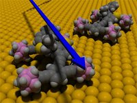
Title: Motorized Nanocar, Credit: Yasuhiro Shirai/Rice UniversityThis animation depicts two motorized nanocars on a gold surface. The nanocar consists of a rigid chassis and four alkyne axles that spin freely
and swivel independently of one another. The wheels are spherical molecules of carbon, hydrogen and boron called p-carborane.

Small Times recognizes Rice chemist for pioneering nanocar research

HOUSTON, Sept. 21, 2006 -- Rice University chemist and nanocar inventor James Tour has been selected Innovator of the Year in Small Times magazine's Best of Small Tech Research Award competition. The awards recognize the best people, products and companies in nanotechnology, microelectromechanical systems (MEMS) and microsystems.

Tour is Chao Professor of Chemistry, professor of mechanical engineering and materials science, professor of computer science and director of the Carbon Nanotechnology Laboratory in Rice's Richard E. Smalley Institute for Nanoscale Science and Technology. He was recognized for his pioneering research in molecular self-assembly, including the development of single-molecule nanocars.

Tour's group unveiled its ultrasmall nanocars in October 2005. Measuring just 3-by-4 nanometers, nanocars have four tires, a rigid chassis and axles that spin freely and swivel independently of one another. About 20,000 nanocars can be parked side-by-side across the diameter of a human hair. The nanocars were imaged in rolling action in collaboration with his colleague Kevin Kelly, assistant professor of electrical and computer engineering.

In announcing the award, Small Times said Tour "is bringing molecular self-assembly to the point of commercial reality. His nanocar is a practical example of molecular manipulation, and his group is hard at work on more sophisticated machines."

Tour designed nanocars as a test system for new methods of molecular self-assembly. During the past year, his research team has extended the original concept, rolling out a motorized nanocar; a nanotruck with a cargo bay; a six-wheeled, three-axled NanoCaterpillar; a nanotrain; a nanobackhoe, complete with flexible extension arm; and an ultrasmall version of the nanocar dubbed the NanoCooper. They are currently working on a high-performance version of the motorized nanocar that contains twin solar-powered motors.

"We want to build things from the bottom-up, one molecule at a time, and in order to do that, we need to transport molecules from place to place," said Tour. "Just as cells use enzymes to assemble proteins and large molecules, we want to design synthetic transporters that are capable of doing much the same thing in non-biological environments." ###

Contact: Jade Boyd jadeboyd@rice.edu 713-348-6778 Rice University

The recipients of the Small Times Magazine Best of Small Tech Awards and runners-up were selected by nominations and applications submitted by companies and individuals. Small Times Media news staff and an industry panel of experts reviewed the applicants and selected one winner and four winners in six categories, plus lifetime achievement. A complete list of winners and runners-up can be found in the November/December issue of Small Times magazine and online at smalltimes.com/.

Technorati Tags: or and or and or and or and or and or and or and or

RELATED: Keywords Nanotech, science Thursday, September 21, 2006 scientists tame tricky carbon nanotubes, Sunday, September 17, 2006 Double Quantum Dots Control Kondo Effect, Saturday, September 16, 2006 Biodegradable napkin, featuring nanofibers, may detect biohazards, Friday, September 08, 2006 Nanoscientists Create Biological Switch from Spinach Molecule, Sunday, September 03, 2006 Sugar metabolism tracked in living plant tissues, in real time, Wednesday, August 30, 2006 'Nanocantilevers' yield surprises critical for designing new detectors, Sunday, August 27, 2006 ion pump cooling hot computer chips, Thursday, August 24, 2006 Honeycomb Network Comprised of Anthraquinone Molecules, Sunday, August 20, 2006 siRNA shrink ovarian cancer tumors, Wednesday, August 16, 2006 Nanotechnology

Thursday, September 21, 2006

scientists tame tricky carbon nanotubes

MIT materials scientists tame tricky carbon nanotubes, Deborah Halber, News Office Correspondent, September 15, 2006

MIT researchers have discovered that certain molecules can attach themselves to metallic carbon nanotubes without interfering with the nanotubes' exceptional ability to conduct electricity. At left, the high conductance state has two molecular orbitals, shown in green. Some molecules even let the nanotube switch between highly conductive, left, and poorly conductive (right, with one red molecular orbital), creating the potential for new applications. Image courtesy / Marzari Lab.Based on a new theory, MIT scientists may be able to manipulate carbon nanotubes --
one of the strongest known materials and one of the trickiest to work with -- without destroying their extraordinary electrical properties.

The work is reported in the Sept. 15 issue of Physical Review Letters, the journal of the American Physical Society.

Carbon nanotubes -- cylindrical carbon molecules 50,000 times thinner than a human hair -- have properties that make them potentially useful in nanotechnology, electronics, optics and reinforcing composite materials. With an internal bonding structure rivaling that of another well-known form of carbon, diamonds, carbon nanotubes are extraordinarily strong and can be highly efficient electrical conductors.

The problem is working with them. There is no reliable way to arrange the tubes into a circuit, partly because growing them can result in a randomly oriented mess resembling a bowl of spaghetti.

Researchers have attached to the side walls of the tiny tubes chemical molecules that work as "handles" that allow the tubes to be assembled and manipulated. But these molecular bonds also change the tubes' structure and destroy their conductivity.

Now Young-Su Lee, an MIT graduate student in materials science and engineering, and Nicola Marzari, an associate professor in the same department, have identified a class of chemical molecules that preserve the metallic properties of carbon nanotubes and their near-perfect ability to conduct electricity with little resistance.

Using these molecules as handles, Marzari and Lee said, could overcome fabrication problems and lend the nanotubes new properties for a host of potential applications as detectors, sensors or components in novel optoelectronics.

Playing with atoms
Marzari and Lee use the fundamental laws of quantum mechanics to simulate material properties that are difficult or impossible to measure, such as molten lava in the Earth's core or atomic motion in a fast chemical reaction. Then they run these simulations on interconnected PCs and use the results to optimize and engineer novel materials such as electrodes for fuel cells and polymers that contract and expand like human muscles.

With the help of a powerful algorithm created by Lee and published last year in Physical Review Letters, the theorists focused on solving some of the problems of working with carbon nanotubes.

Like fuzzy balls and Velcro, the hexagon of carbon that makes up a nanotube has a predilection for clinging to other hexagons. One of the many challenges of working with the infinitesimally small tubes is that they tend to stick to each other.

Attaching a molecule to the sidewall of the tube serves a double purpose: It stops nanotubes from sticking so they can be processed and manipulated more easily, and it allows researchers to control and change the tubes' electronic properties. Still, most such molecules also destroy the tubes' conductance because they make the tube structurally more similar to a diamond, which is an insulator, rather than to graphite, a semi-metal.

Lee and Marzari used Lee's algorithm to identify a class of "molecular handles" (carbenes and nitrenes) that stop this from happening and preserve the tubes' original conductivity. "We now have a way to attach molecules that allows us to manipulate the nanotubes without losing their conductance," Marzari said.

Carbenes and nitrenes work by breaking a molecular bond on the nanotube's wall while creating their own new bond to the tube. This process -- one bond formed, one bond lost -- restores the perfect number of bonds each carbon atom had in the original tube and "conductance is recovered," Marzari said.

Some molecular handles can even transform between a bond-broken and a bond-intact state, allowing the nanotubes to act like switches that can be turned on or off in the presence of certain substances or with a laser beam. "This direct control of conductance may lead to novel strategies for the manipulation and assembly of nanotubes in metallic interconnects, or to sensing or imaging devices that respond in real-time to optical or chemical stimuli," Marzari said.

The next step is for experiments to confirm that the approach works.

This work is supported by the MIT Institute for Soldier Nanotechnologies and the National Science Foundation

A version of this article appeared in MIT Tech Talk on September 20, 2006 (In PDF Format).

Technorati Tags: or and or and or and or and or and or and or and or

RELATED: Keywords Nanotech, science Sunday, September 17, 2006 Double Quantum Dots Control Kondo Effect, Saturday, September 16, 2006 Biodegradable napkin, featuring nanofibers, may detect biohazards, Friday, September 08, 2006 Nanoscientists Create Biological Switch from Spinach Molecule, Sunday, September 03, 2006 Sugar metabolism tracked in living plant tissues, in real time, Wednesday, August 30, 2006 'Nanocantilevers' yield surprises critical for designing new detectors, Sunday, August 27, 2006 ion pump cooling hot computer chips, Thursday, August 24, 2006 Honeycomb Network Comprised of Anthraquinone Molecules, Sunday, August 20, 2006 siRNA shrink ovarian cancer tumors, Wednesday, August 16, 2006 Nanotechnology

Sunday, September 17, 2006

Double Quantum Dots Control Kondo Effect

Nano News: Double Quantum Dots Control Kondo Effect, Thursday Sep 14, 2006 by ANDREA GIBSON

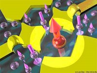
Nanoscale metallic electrodes (in yellow) can be used to confine electrons in small regions, forming quantum dots. Two quantum dots connected to each other form a double quantum dot. In this case, one of the dots is in the Kondo state, in which the magnetic moment of the confined electron (large red arrow) is compensated ('screened') by the magnetic moment of surrounding electrons, resulting in a zero net magnetic moment for the entire system. art by: Luis Dias/Ohio UniversityNanoscale metallic electrodes (in yellow) can be used to confine electrons in small regions, forming quantum dots. Two quantum dots connected to each other form a double quantum dot.
In this case, one of the dots is in the Kondo state, in which the magnetic moment of the confined electron (large red arrow) is compensated (“screened”) by the magnetic moment of surrounding electrons, resulting in a zero net magnetic moment for the entire system. art by: Luis Dias/Ohio University

ATHENS, Ohio — Two quantum dots connected by wires could help scientists better control the Kondo effect in experiments, according to a study by Ohio University and University of Florida physicists published in a recent issue of Physical Review Letters.

The Kondo effect occurs when electrons become trapped around the magnetic impurities in semiconductor materials, which prompts the electrons to change their spin. This phenomenon has intrigued scientists, as electronic correlations can create interesting and complex behavior in materials.

In the new work, scientists demonstrate how the two quantum dot system can behave in two different and interesting ways: As a simile for a Kondo-effect system where one quantum dot is used to "filter" the effect of the current leads, and as a way to study "pseudo-gapped" systems and correlations in them, which can help scientists understand structures such as superconductors.

“This last part is of great current interest to theorists and experimentalists who are exploring what are called quantum phase transitions, which are changes in systems that alter their behavior dramatically as a function of some parameter while remaining at zero (or very low) temperature,” said Sergio Ulloa, a professor of physics and astronomy at Ohio University.

The study, funded by the National Science Foundation, was conducted by Luis Dias da Silva, Nancy Sandler and Ulloa, all members of the Ohio University’s Nanoscale and Quantum Phenomena Institute, and Kevin Ingersent of the University of Florida.

Technorati Tags: or and or and or and or and or and or and or and or

RELATED: Keywords Nanotech, science Saturday, September 16, 2006 Biodegradable napkin, featuring nanofibers, may detect biohazards, Friday, September 08, 2006 Nanoscientists Create Biological Switch from Spinach Molecule, Sunday, September 03, 2006 Sugar metabolism tracked in living plant tissues, in real time, Wednesday, August 30, 2006 'Nanocantilevers' yield surprises critical for designing new detectors, Sunday, August 27, 2006 ion pump cooling hot computer chips, Thursday, August 24, 2006 Honeycomb Network Comprised of Anthraquinone Molecules, Sunday, August 20, 2006 siRNA shrink ovarian cancer tumors, Wednesday, August 16, 2006 Nanotechnology

Saturday, September 16, 2006

Biodegradable napkin, featuring nanofibers, may detect biohazards

CU biodegradable wipe would quickly detect biohazards, from avian flu to E. coli, By Susan Lang

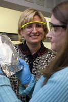
Jamie Mullally '07, right, a Cornell Presidential Research Scholar, and Margaret Frey, assistant professor of textiles and apparel, examine a nonwoven nanofiber fabric on aluminum foil backing. Mullally will complete an honors thesis on the biorecognition fabrics in spring '07. Copyright © Cornell UniversityJamie Mullally '07, right, a Cornell Presidential Research Scholar, and Margaret Frey, assistant professor of textiles and apparel, examine a nonwoven nanofiber fabric on aluminum foil backing.
Mullally will complete an honors thesis on the biorecognition fabrics in spring '07. Copyright © Cornell University.

Detecting bacteria, viruses and other dangerous substances in hospitals, airplanes and other commonly contaminated places could soon be as easy as wiping a napkin or paper towel across a surface

"It's very inexpensive, it wouldn't require that someone be highly trained to use it, and it could be activated for whatever you want to find," said Margaret Frey, the Lois and Mel Tukman Assistant Professor of Fiber Science and Apparel Design at Cornell University. "So if you're working in a meat-packing plant, for instance, you could swipe it across some hamburger and quickly and easily detect E. coli bacteria." She reported on the research Sept. 11 at the American Chemical Society's national meeting.

Once fully developed, the biodegradable absorbent wipe would contain nanofibers containing antibodies to numerous biohazards and chemicals and would signal by changing color or through another effect when the antibodies attached to their targets. Users would simply wipe the napkin across a surface; if a biohazard were detected, the surface could be disinfected and retested with another napkin to be sure it was no longer contaminated.

In work conducted with Yong Joo, assistant professor of chemical and biomolecular engineering, and Antje Baeumner, associate professor of biological and environmental engineering, both at Cornell, Frey developed nanofibers with platforms made of biotin, a part of the B vitamin complex, and the protein streptavidin, which can hold the antibodies. Composed of a polymer compound made from corn, the nanofibers could be incorporated into conventional paper products to keep costs low. Nanofibers, with diameters near 100 nanometers (a nanometer is one-billionth of a meter, or about three times the diameter of an atom), provide extremely large surface areas for sensing and increased absorbency compared with conventional fibers.

"The fabric basically acts as a sponge that you can use to dip in a liquid or wipe across a surface," Frey said. "The fabric itself will transport and concentrate the targeted biohazard. As you do that, antibodies in the fabric are going to selectively latch onto whatever pathogen that they match. Using this method we should, in theory, be able to quickly activate the fabric to detect whatever is the hazard of the week, whether it is bird flu, mad cow disease or anthrax."

Frey and her colleagues are still working on ways, such as a color change, for the fabric to signal that it has identified the contaminant.

"We're probably still a few years away from having this ready for the real world," Frey said, "but I really believe there is a place for this type of product that can be used by people with limited training to provide a fast indication of whether a biohazard is present."

This research was supported by the National Research Initiative of the U.S. Department of Agriculture's Cooperative State Research, Education and Extension Service.

### Contact: Blaine Friedlander bpf2@cornell.edu 607-254-8093 Cornell University News Service

Technorati Tags: or and or and or and or and or and or and or and or

RELATED: Keywords Nanotech, science Friday, September 08, 2006 Nanoscientists Create Biological Switch from Spinach Molecule, Sunday, September 03, 2006 Sugar metabolism tracked in living plant tissues, in real time, Wednesday, August 30, 2006 'Nanocantilevers' yield surprises critical for designing new detectors, Sunday, August 27, 2006 ion pump cooling hot computer chips, Thursday, August 24, 2006 Honeycomb Network Comprised of Anthraquinone Molecules, Sunday, August 20, 2006 siRNA shrink ovarian cancer tumors, Wednesday, August 16, 2006 Nanotechnology

Friday, September 08, 2006

Nanoscientists Create Biological Switch from Spinach Molecule

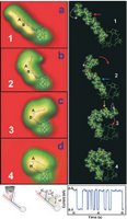
Scientists used a scanning tunneling microscope to manipulate chlorophyll-a into four positions. art by: Saw-Wai HlaNanoscientists Create Biological Switch from Spinach Molecule, Scientists used a scanning tunneling microscope to manipulate chlorophyll-a into four positions. art by: Saw-Wai Hla, Tuesday Sep 05, 2006, by Andrea Gibson
ATHENS, Ohio – Nanoscientists have transformed a molecule of chlorophyll-a from spinach into a complex biological switch that has possible future applications for green energy, technology and medicine.

The study offers the first detailed image of chloropyhll-a – the main ingredient in the photosynthesis process – and shows how scientists can use new technology to manipulate the configuration of the spinach molecule in four different arrangements, report Ohio University physicists Saw-Wai Hla and Violeta Iancu in today’s early edition of the journal Proceedings of the National Academy of Sciences.

The scientists used a scanning tunneling microscope to image chlorophyll-a and then injected it with a single electron to manipulate the molecule into four positions, ranging from straight to curved, at varying speeds. (View a movie here) Though the Ohio University team and others have created two-step molecule switches using scanning tunneling microscope manipulation in the past, the new experiment yields a more complex multi-step switch on the largest organic molecule to date.

The work has immediate implications for basic science research, as the configuration of molecules and proteins impacts biological functions. The study also suggests a novel route for creating nanoscale logic circuits or mechanical switches for future medical, computer technology or green energy applications, said Hla, an associate professor of physics.

“It’s important to understand something about the chlorophyll-a molecule for origin of life and solar energy conversion issues,” he said.

The study was funded by Ohio University’s Nanobiotechnology Initiative and the U.S. Department of Energy. Hla is a member of the university’s Quantitative Biology Institute and Nanoscale & Quantum Phenomena Institute. Iancu is a doctoral candidate in the Department of Physics and Astronomy.

Additional information, Contact: Saw-Wai Hla, (740) 593-1727, hla@ohio.edu
Proceedings of the National Academy of Sciences link to article
Department of Physics and Astronomy

Related News:
Scientists Create the First Synthetic Nanoscale Fractal Molecule

Technorati Tags: or and or and or and or and or and or and or and or

RELATED: Keywords Nanotech, science Sunday, September 03, 2006 Sugar metabolism tracked in living plant tissues, in real time, Wednesday, August 30, 2006 'Nanocantilevers' yield surprises critical for designing new detectors, Sunday, August 27, 2006 ion pump cooling hot computer chips, Thursday, August 24, 2006 Honeycomb Network Comprised of Anthraquinone Molecules, Sunday, August 20, 2006 siRNA shrink ovarian cancer tumors, Wednesday, August 16, 2006 Nanotechnology

Sunday, September 03, 2006

Sugar metabolism tracked in living plant tissues, in real time

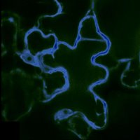
This false-color image shows a cell from the epidermis of an Arabidopsis thaliana plant; the cell has been marked with fluorescent imaging sensors designed to detect the sugar glucose. In this image, only the densely packed interior of the cell in which most metabolic functions occur—called the cytosol—is targeted by the glucose sensors. The dark area sits inside the vacuole—a large storage organelle that can occupy up to 90% of the cell’s volume. (Image courtesy Sylvie Lalonde and Wolf Frommer;)This false-color image shows a cell from the epidermis of an Arabidopsis thaliana plant; the cell has been marked with fluorescent imaging sensors designed to detect the sugar glucose.
In this image, only the densely packed interior of the cell in which most metabolic functions occur—called the cytosol—is targeted by the glucose sensors. The dark area sits inside the vacuole—a large storage organelle that can occupy up to 90% of the cell’s volume. (Image courtesy Sylvie Lalonde and Wolf Frommer; click for higher resolution.)

Stanford, CA – Scientists at Carnegie’s Department of Plant Biology have made the first real-time observations of sugars in the cells of intact and living plant tissues. With the help of groundbreaking imaging techniques, the group has determined that plants maintain extremely low levels of sugar in their roots—as much as 100,000 times lower than previous estimates. The new technology will enable new studies of sugar metabolism in plants, which will inform the effort to engineer higher crop yields for food and biofuel production.

Led by Carnegie staff member Wolf Frommer, the researchers designed genetically-encoded fluorescent tags to monitor glucose, an important sugar, in leaf and root tissues of the model plant Arabidopsis thaliana. The technique has allowed the researchers to track glucose over time and space at unprecedented detail, in living and undisturbed plant tissues. The work appears in the September issue of the journal Plant Cell*. The group has also developed a FRET sensor for sucrose, a major transport sugar in plants. This work will appear in the September issue of the Journal of Biological Chemistry**.

“Until now, we have had few clues regarding how much sugar is in an individual cell in a multicellular plant,” Frommer said. “We normally grind up a leaf or a root and average the information for all cells, but if sugar levels rise in one cell and drop in another, we would see no change in this average.” Also, because the cell can distribute sugar among subcellular organelles, it is nearly impossible to know how much sugar is in any cell compartment at a given time.
“Time resolution is another problem,” Frommer added. “We can sample tissue at intervals, but if the sugar changes in waves, we might miss the right time point. Our new technology addresses all of these problems by measuring sugar flux in real time in individual cells, with subcellular resolution.”

Frommer and his colleagues have used similar imaging tags, called fluorescent resonance energy transfer (FRET) sensors, to track sugars and neurotransmitters in animal cells. Most recently, the group used FRET sensors to study glutamate, an important mammalian neurotransmitter. Frommer has tracked glucose in cultured mammalian cells, but until now, plant tissues had proven problematic because of interference from the plants’ virus defense mechanisms, as well as high background fluorescence in some plants.

To surmount these issues, Frommer’s team dramatically improved the sensors, while inserting them in mutant Arabidopsis plants with disabled defense genes. The fluorescent tags worked well where they had failed before.

“It may not be ideal to use defense-mutant plants—the ideal would be for the sensors to work in any wild-type genetic background,” Frommer explained. “But proving that the sensors can work in plants is an important first step. Now we can begin addressing important questions about the way plants manage sugar distribution while we continue to improve the sensors.”

In preliminary experiments, Frommer’s group compared fluctuations in glucose levels in root tissue and leaf epidermis—the topmost layer that absorbs sunlight—and found that the plant maintained glucose at higher levels in leaf tissue than in roots. In fact, the researchers found that root cells contain sugar at concentrations at least 100,000 times lower than previous estimates.

FRET sensors are encoded by genes that, in theory, can be engineered into any cell line or organism. They are made of two fluorescent proteins that produce different colors of light—one cyan and one yellow—connected by a third protein that resembles a hinged clam shell. The two fluorescent proteins are derived from jellyfish, and the third from a bacterium; the shape of the clam shell protein determines which sugar or other molecule the sensor can detect. When a target molecule such as glucose or sucrose binds to the third protein, the hinge opens, changing the distance and orientation of the fluorescent proteins. This physical change affects the energy transfer between the cyan and yellow markers.

When the researchers hit the tags with light of a specific wavelength, the cyan tag starts to fluoresce. If the yellow tag is close enough, the cyan tag will transfer its energy to the yellow tag, causing it to resonate and fluoresce as well. This energy transfer affects how much cyan and yellow fluorescence can be seen, and by calculating this ratio, researchers can accurately track molecules such as glucose and sucrose in both time and space.

“The strength of this technology lies in its elegant simplicity; with the power of computational design, we can potentially design FRET tags to detect virtually any small molecule in living cells,” Frommer said. “Imaging techniques like this are the next frontier in the study of metabolism, and will help to answer some of the most pressing questions on plant biologists’ minds, such as the role of individual genes in the distribution of sugars. This in turn can help us engineer plants to produce more biomass.”

Carnegie Contact: Dr. Wolf Frommer; wfrommer@stanford.edu or (650) 325-1521 x208, News ReleaseThursday, August 31, 2006

*For a copy of the Plant Cell paper, go to: plantcell.org/cgi/content/short/

**For a copy of the Journal of Biological Chemistry paper, go to: jbc.org/cgi/reprint/

The two projects were supported by grants from the U.S. Department of Energy, the National Institutes of Health, and the European Science award of the Körber Foundation, Hamburg.

The Carnegie Institution of Washington (carnegieinstitution.org) has been a pioneering force in basic scientific research since 1902. It is a private, nonprofit organization with six research departments throughout the U.S. Carnegie scientists are leaders in plant biology, developmental biology, astronomy, materials science, global ecology, and Earth and planetary science. The Department of Plant Biology is located at 260 Panama St., Stanford, CA 94305

Technorati Tags: or and or and or and or and or and or and or and or

RELATED: Keywords Nanotech, science Wednesday, August 30, 2006 'Nanocantilevers' yield surprises critical for designing new detectors, Sunday, August 27, 2006 ion pump cooling hot computer chips, Thursday, August 24, 2006 Honeycomb Network Comprised of Anthraquinone Molecules, Sunday, August 20, 2006 siRNA shrink ovarian cancer tumors, Wednesday, August 16, 2006 Nanotechnology